
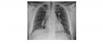
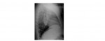
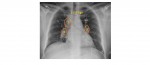
The following chest x-rays are from a 35 year old man who has presented with chest pain and exertional dyspnoea. What radiological sign is noticeable in these x-rays?
[peekaboo_link name=”Answer”]Answer[/peekaboo_link] [peekaboo_content name=”Answer”]
The PA chest x-ray shows enlargement of the right paratracheal, right and left hilar lymph nodes. This pattern of lymph node enlargement is suggestive of sarcoidosis. The radiological sign visible is the 1-2-3 sign or the Garland’s triad.
This patient had a CT chest scan and a lymph node biopsy and was subsequently diagnosed with stage II sarcoidosis.
Staging of Sarcoidosis:
Stage 1 – Bilateral hilar lymphadenopathy
Stage 2 – Bilateral hilar adenopathy and infiltrates
Stage 3 – Infiltrates alone
Stage 4 – Fibrosis
Sarcoidosis is a chronic granulomatous disease of unknown aetiology. Though it most commonly affects the lungs, it can technically manifest in any organ within the body.
Organs affected are:
- Lungs (90%) – hilar adenopathy, lung infiltrates, pulmonary fibrosis, pulmonary hypertension.
- Liver (60-90%) – abnormal LFTs.
- Spleen (40%) – splenomegaly, anaemia.
- Eyes (30%) – uveitis, keratoconjunctivitis.
- Skin (30%) – lupus pernio, erythema nodosum.
- Joint (25%) – acute or chronic arthritis.
- Endocrine (10%) – hypercalcaemia.
- CNS (5%) – cranial nerve palsies (commonly facial nerve), aseptic meningitis.
- CVS (5%) – conduction defects, heart block, cor pulmonale.
Reference: http://emedicine.medscape.com/article/809047
[/peekaboo_content]