
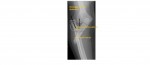
The following right forearm x-rays are from a 16 year old who is experiencing forearm swelling and deformity after a fall on outstretched hand. What can you note on the x-rays?
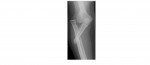
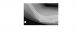

The x-rays show a Monteggia fracture dislocation; this is a fracture of the ulnar shaft at the junction of the upper and mid-third with a radial head dislocation.
The patient underwent operative reduction of the radial head with internal fixation of the ulnar shaft fracture.
The forearm bones are strongly connected to each other by ligaments and it is difficult to fracture only one of the bones. However, if only one bone appears fractured, a covert injury of the other bone is likely and this usually manifests as a dislocation of the non-fractured bone.
If a long bone fracture is suspected, it is important to always x-ray the joint above and below.
A radio-capitellar line is useful to detect a radial head dislocation. A line drawn along the mid-shaft of the radius should pass through the capitellum on the AP as well as the lateral view. If not, the radial head is dislocated.
Reference: Grainger and Allison’s Diagnostic Radiology
[/peekaboo_content]