
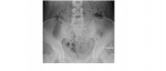
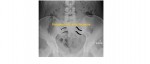
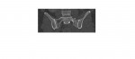
The following pelvic x-ray is from a 40 year old who has been involved in a motor vehicle accident. What 3 fractures can you note on the x-ray?
[peekaboo_link name=”Answer”]Answer[/peekaboo_link] [peekaboo_content name=”Answer”]
The 3 fractures seen are:
1. Left sacral alar fracture – there is disruption of the sacral foramina, which appear as curved white lines (arcuate lines).
2. Right superior pubic ramus fracture.
3. Transverse process fracture of L1 on the left (sorry but it is difficult to appreciate on the given film unless magnified).

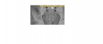
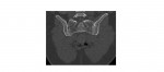
CT scans from the same patient which clearly show the left sacral alar fracture:
The sacrum is formed by the fusion of 5 sacral vertebrae; it contains 4 foramina which transmit sacral nerve roots (S1-S4). S1 and S2 roots carry the highest risk of injury.
Sacral fractures fall into 3 categories based on the Denis classification:
- Zone 1 – Fractures lateral to the sacral foramina; L5 root can be injured.
- Zone 2 – Fractures through the foramina; unilateral sacral anaesthesia (unilateral preservation of nerves is enough for bowel and bladder control).
- Zone 3 – Fractures medial to the foramina into the spinal canal; risk to the cauda equina.
Sacral alar fractures are difficult to diagnose and commonly missed because of the overlying bowel gas. The arcuate lines, which normally appear as smooth curved white lines, need to be carefully followed and compared with the opposite side; asymmetry/discontinuity/disruption is indicative of sacral fracture. CT scan is the diagnostic test.
Reference – http://www.orthobullets.com/trauma/1032/sacral-fractures
[/peekaboo_content]