
The following erect chest and abdominal x-rays are from a 60 year old with sudden onset of abdominal pain. What can you deduce from these images?

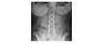
The cardiomediastinal contour appears normal. There is left basal atelectasis. There is also a triangular pocket of air which can be seen below the left hemi diaphragm.
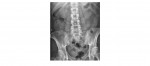
The abdominal x-rays do not show any features of bowel obstruction. The triangular area of gas below the left hemi diaphragm can again be seen on the erect abdominal x-ray.
Is this pneumoperitoneum?
The patient went onto have a CT scan of the abdomen which showed a perforated gastric ulcer that was causing pneumoperitoneum.

Although mostly signs of pneumoperitoneum appear on the right side, perhaps because of pre-existing adhesions, gas may accumulate on the left side either lateral to the stomach or between the gastric air bubble and the left hemi diaphragm. And as such, this area needs to be carefully scrutinised.
Reference: Dynamic radiology of the Abdomen: Morton Meyers
[/peekaboo_content]