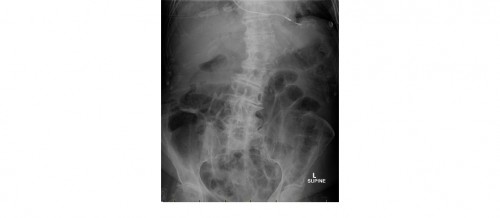
It is 3am and your next patient is a 70 year old female with abdominal pain and vomiting. To make things complicated, she also gives a history of runny stools. Her supine abdominal x-ray is as follows.
You are not too sure of what is going on and happen to notice your inpatient colleague sitting next to you and ask him for his opinion. He is not too impressed with the film and you are almost certain that he is going to refuse your referral. Would you refer this elderly patient with abdominal pain and vomiting to him anyway? What can you see on the x-ray?
[peekaboo_link name=”Answer”]Answer[/peekaboo_link] [peekaboo_content name=”Answer”]The supine abdominal x-ray is incomplete as you cannot see the top of the right diaphragm and the lower part of the abdomen. There is small bowel dilatation in the lower abdomen. There is faecal loading in the right colon. The large bowel segments in the upper abdomen look nondilated.There are calcified costal cartilages bilaterally. There is a single lead pacemaker wire visible.
What else can also be seen on this x-ray?
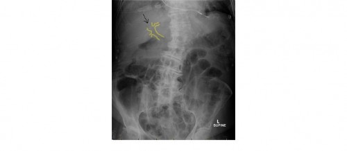
Air where it should not be!
There is pneumobilia which is air in the biliary tree. Air in the biliary tree in the context of small bowel obstruction indicates small bowel obstruction secondary to a gallstone. In the given x-ray there is no gallstone identified on the x-ray; in other words, there is no Rigler’s triad.
Rigler’s triad is the presence of pneumobilia, small bowel obstruction and impacted gall stone seen in the distal ileum at the ileocaecal valve.
What are the causes of pneumobilia ?
- following passage of a gallstone
- recent sphincterotomy or ERCP
- emphysematous cholecystitis (in diabetics)
- lax sphincter of Oddi
- previous Whipple’s surgery
What is the other important differential when you see gas in the liver area?
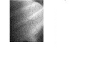
Portal venous gas: gas in the portal venous system due to ischaemic bowel, portal pyaemia etc. It is a much more serious condition. Portal venous gas tends to reach the periphery of the hepatic shadow whereas pneumobilia tends to be central and does not extend to the periphery of liver and is typically seen in the right paravertebral area (left liver lobe area).
(Portal venous gas image, courtesy of emedicine.medscape.com)
Gallstone ileus is seen in less than 1-3 % of patients with cholelithiasis. The incidence of pneumobilia in gallstone ileus is about 70 %.
And finally, presence of diarrhoea or frequest bowel action is an early feature in small bowel obstruction due to GI emptying phenomenon. Patients should not be mistaken as having ‘gastro’.
[/peekaboo_content]


