
This lumbar spine AP x-ray is from a 52 year old chronic alcoholic with low back pain. What incidental finding can you observe on the x-ray?

The x-ray does not show any abnormality of the lumbar spine. There are multiple small specks of calcification projected over the right side of the L2 vertebral body and transverse process, anatomically located over the region of pancreas, and this is consistent with calcified pancreas.
Anatomically, pancreas lies across L1-L2.
 Pancreatic calcifications have largely been associated with alcoholic chronic pancreatitis. Chronic pancreatitis that is caused by hyperparathyroidism, tropical pancreatitis and idiopathic pancreatitis may also result in intraductal calculi (reference: http://www.ajronline.org/doi/full/10.2214/ajr.178.1.1780079).
Here are some cases of incidental calcifications noted on abdominal x-rays.
1. Cluster of gall bladder calculi on a lumbar spine x-ray (thanks to Dr. Ignatius Munjodzi for this image):
 2. Single gall bladder calculus:
 3. Stag horn calculus in the left kidney:
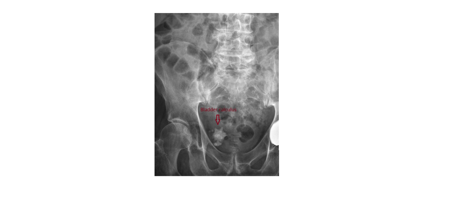
 4. Urinary bladder calculus (suspicion of an associated TCC of bladder on CT):
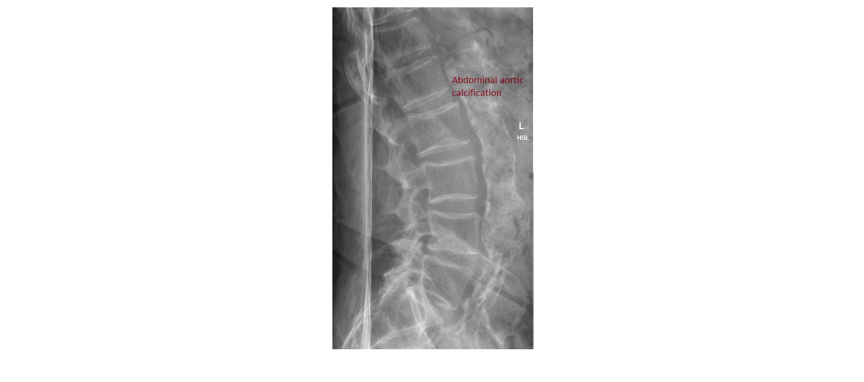
 5. Patient with low back pain, stable haemodynamics. The plain x-ray below shows a heavily calcified and dilated aorta. A CT conducted soon after the x-ray showed an abdominal aortic aneurysm with a maximal diameter of 4cm which was just proximal to the aortic bifurcation with no leak.
excelent cases