
A 52 year old female presents to you with a history of constant low back pain which has been going on for about 3 weeks. She has been previously well and is a nonsmoker. There is no history of trauma. You want to request an x-ray of her lumbar spine but, at the same time, you are hesitant because you have been criticised by your consultant a few times for requesting imaging for low backpain patients without red flags. You sneakily get one done anyway when your consultant is busy in resus and here is the x-ray. What can you see?
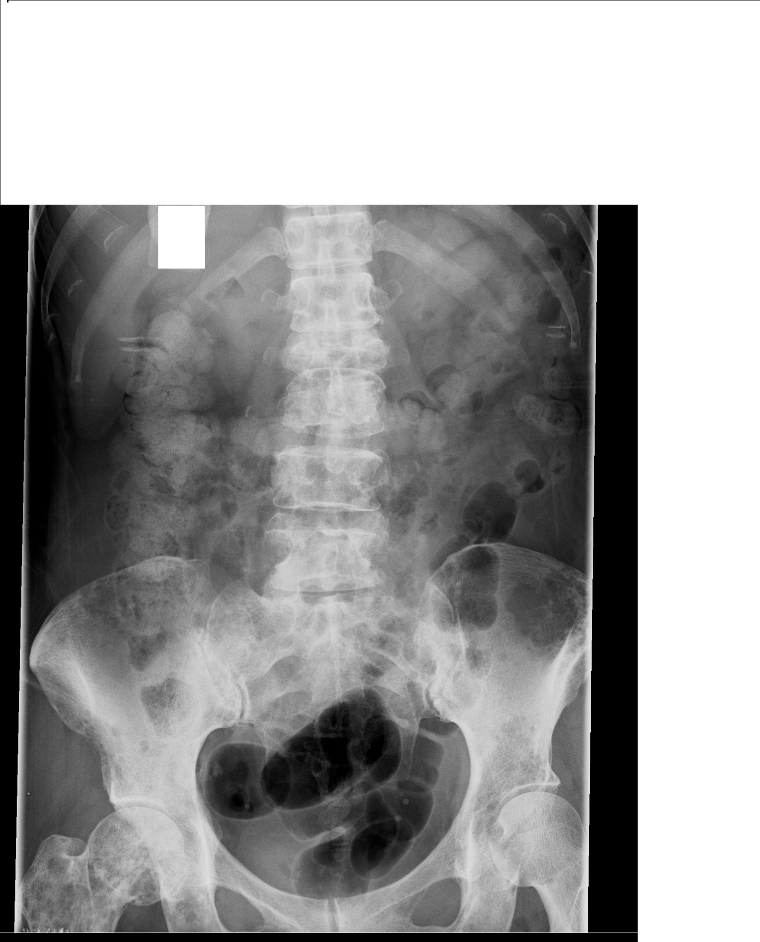
The x-ray shows a collapse of L2 with 60 -70% reduction in height with destruction pedicles from L2-L5. There is a large lytic area in the left iliac wing. There are patchy lytic and sclerotic areas in the right acetabulum, femoral neck and greater trochanter area. There are lytic lesions in the left acetabulum. To sum up, there are multiple skeletal mets.
What are the common primaries that metastasize to bone ? Breast, lung, prostate, kidney and thyroid. Possibility of haematological malignancy should also be kept in mind. In this patient, the most likely site is breast but further investigations are needed for confirmation.
In regards to obtaining images in low backpain patients, you should largely be guided by the presence of ‘red flags’. The diagnostic yield in the absence of red flags is low. The red flags are
- Recent major trauma
- Mild trauma if age > 50
- Fever
- History of malignancy or immune compromise
- Unexplained weight loss
- IVDU
- Osteoporosis or steroid use
- Age > 70
- Suspicion of ankylosisng spondylosis
- Suspected cauda equina syndrome
(information on red flags courtesy www.imagingpathways.health.wa.gov.au )
Back pain that is nocturnal, not relieved with bed rest and thoracic back pain should also be viewed as red flags.
[/peekaboo_content]