
The following skull x-ray is from a 3 year old with a history of a head injury 72 hours prior to presentation. He had a fall in the playground with no LOC and one incidence of vomiting post-injury. After suffering the injury, he had been observed in the ED for 4 hours, remained well and was then discharged home. He has now been brought in by his parents as he’s been irritable since the initial discharge. On examination, the only finding is a boggy swelling in the right parietal area. A skull x-ray was performed by the treating physician; what can be observed?
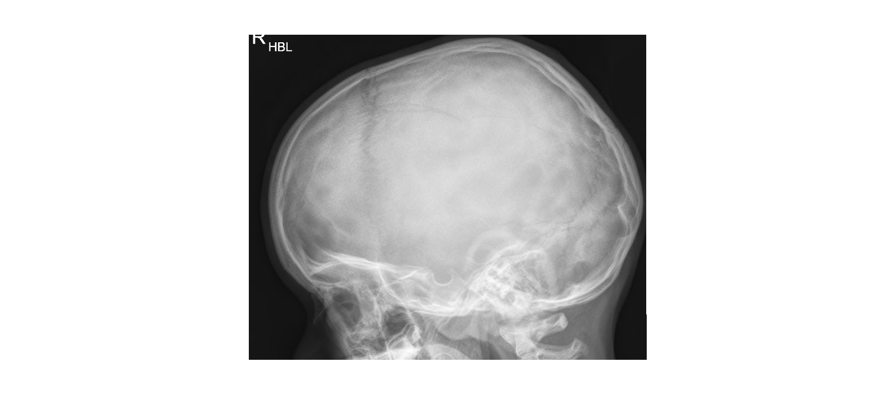
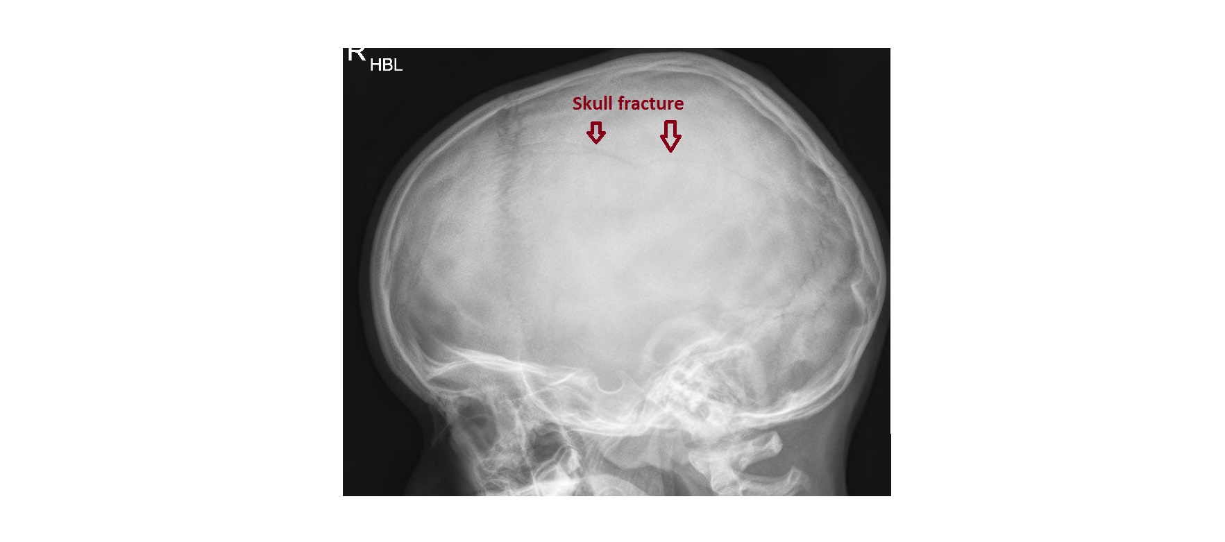
The skull x-ray shows a linear lucency over the parietal bone, consistent with a fracture. A skull fracture is associated with an increased incidence of an intracranial bleed. The child underwent a CT scan of the head which confirmed the fracture and did not show any intracranial bleed.

This child was admitted under the care of a neurosurgeon and did well.
How to differentiate a skull fracture from a suture line? A skull fracture appears as a linear lucency with non-sclerotic edges. In contrast, sutures show a zig-zag pattern and sclerotic borders.
Caveat: absence of skull fracture on a plain film does not rule out a significant intracranial injury.
Thanks to Dr. Michael Lovegrove for the case and images.
Reference: Fundamentals of Diagnostic Radiology by Brant and Helms
[/peekaboo_content]