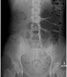
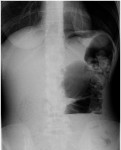
It is 2 am and you are about to see a 19 year old female patient with abdominal pain. As you approach her cubicle, you can hear her screaming. You find the patient holding her abdomen and crying out in pain.
She is holding a vomit bag in her other hand and will not talk to you when you want to know more details about her presentation. You briefly examine her abdomen and it feels soft but slightly distended and generally tender. You are convinced that there is nothing serious going on.
However, you decide to obtain an x-ray of her abdomen, not expecting to find much in terms of findings. Here are her x-rays. What do they show?
[peekaboo_link name=”Answer”]Answer[/peekaboo_link] [peekaboo_content name=”Answer”]The abdominal x-ray shows significantly distended large bowel loop (haustra clearly visible) which has crossed the midline towards left upper quadrant. When you measure it, it is approximately 9.5 cm in diameter. There is faecal material within the loop as well as in the rectum and right side of colon. This patient has caecal volvulus.
Caecal volvulus accounts for 25-40% of all cases of colonic volvulus. Usually secondary to abnormal mesenteric fixation and excessive motility. Can occur following a colonoscopy, secondary to adhesions, constipation and in pregnancy. Marathon runners generally have a higher incidence of this.
Common age group is 20-40 years as opposed to sigmoid volvulus which affects the elderly. Caecal volvulus should be suspected in younger population with bowel obstruction without known risk factors.
The distended caecum can be seen on the x-ray, pointing to the LUQ (usually) but it can lie anywhere including the pelvis. However, in sigloid volvulus, the twisted sigmoid most commonly lies in the RUQ.
There are 2 types:
- Majority (90%) due to axial rotation causing twisting of the mesentery and blood vessels.
- 10% is due to caecal bascule due to the folding of the caecum and ascending colon in a cephalad direction.
On plain x-rays, there may be an inverted U appearance of distended caecum with coffee bean sign as a result of midline crease corresponding to mesenteric root in the distended caecum.
Caecal volvulus is a surgical emergency. Caecal diameter > 10-12 cm may indicate impending perforation.
For those interested, here is a case report on caecal volvulus as an unusual presentation.
[/peekaboo_content]

