
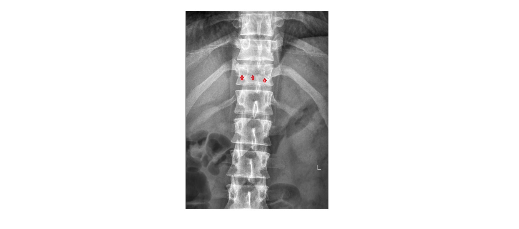
The frontal thoracolumbar x-ray shows a horizontal lucency through the vertebral body and pedicles of the T11 vertebra. There is a subtle reduction in the height of the vertebra.
The lateral (HBL) view shows a fracture of T11 involving the anterior, middle and posterior vertebral columns. There is about 20% compression of the anterior vertebra.
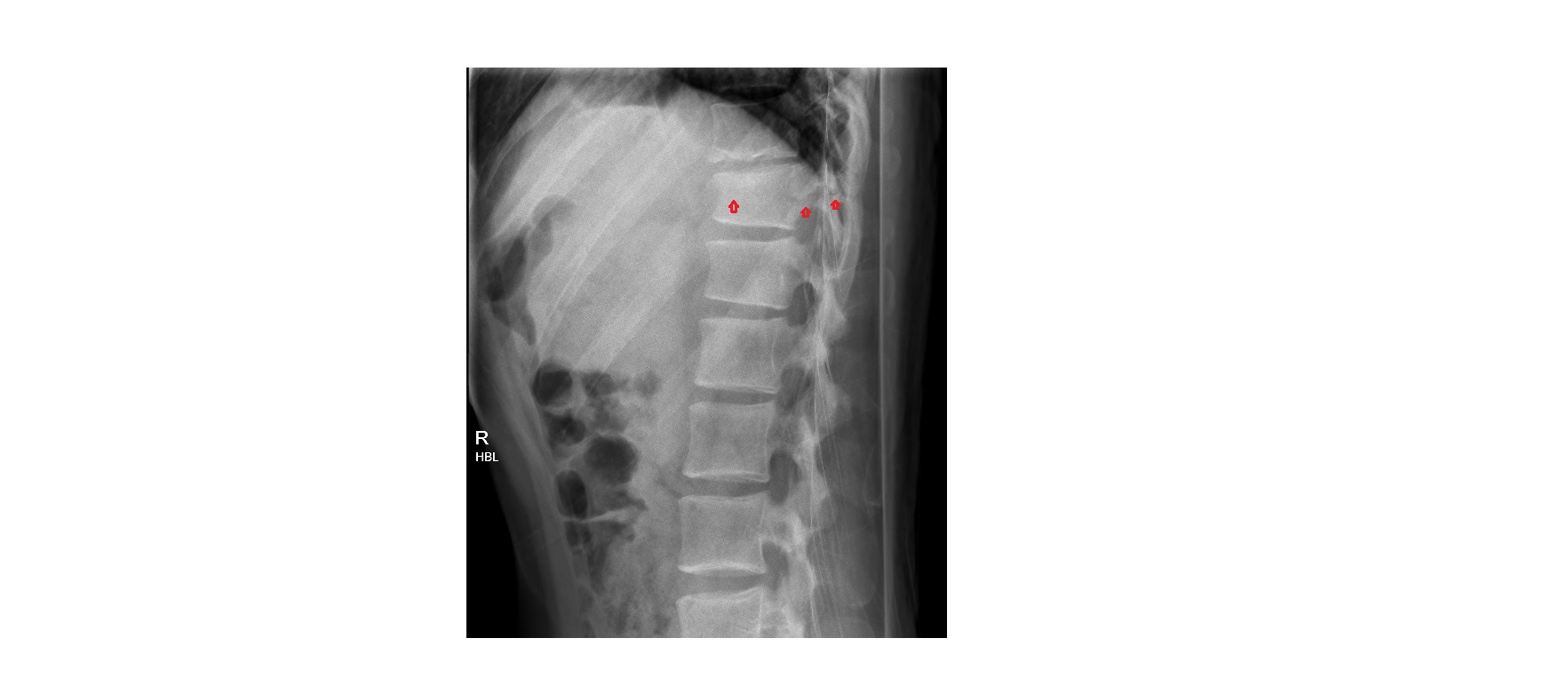 This is a Chance fracture, which is an unstable injury involving all three columns of the vertebral body (anterior, middle and posterior).
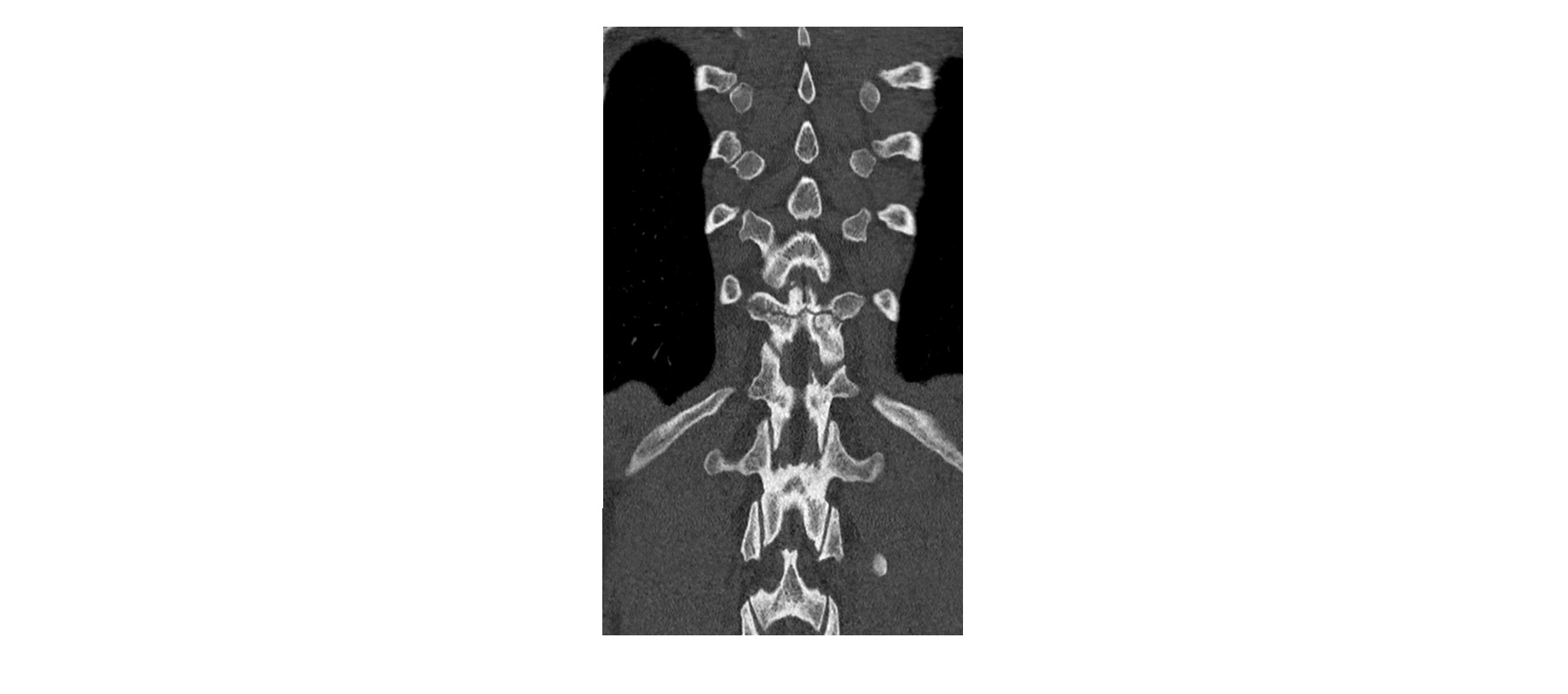
Here are the coronal and sagittal CT images from the same patient:

Chance fractures occur as a result of acute hyper-flexion and distraction injury to the spine around the thoracolumbar area, especially T11 to L2. This is due to the relative immobility of the thoracic spine as compared to the lumbar spine.
Common mechanism is MVA due to incorrectly worn lap seat-belt. It is also less commonly caused by a fall from a height.
This is an unstable injury but neurological complications are infrequent. However, these injuries often have a high association with intra-abdominal and retroperitoneal injuries (up to 50-60%).
Further details can be found here.