
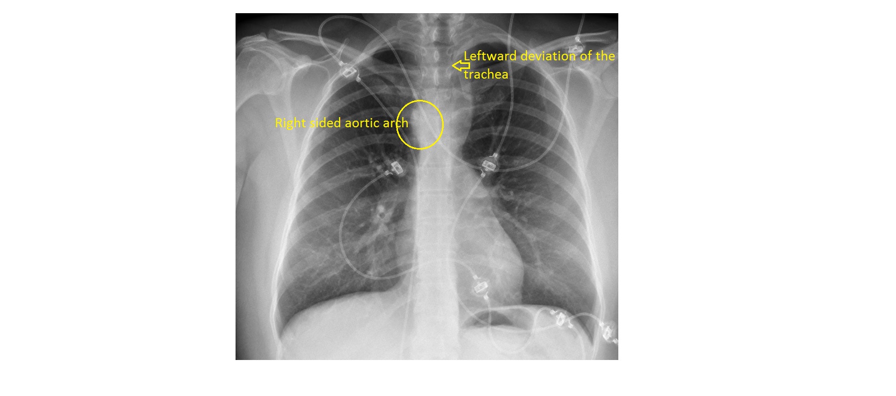
The frontal chest x-ray shows an abnormal superior mediastinum. The aortic knob is to the right of trachea and the trachea is mildly displaced to the left, suggestive of a right sided aortic arch.
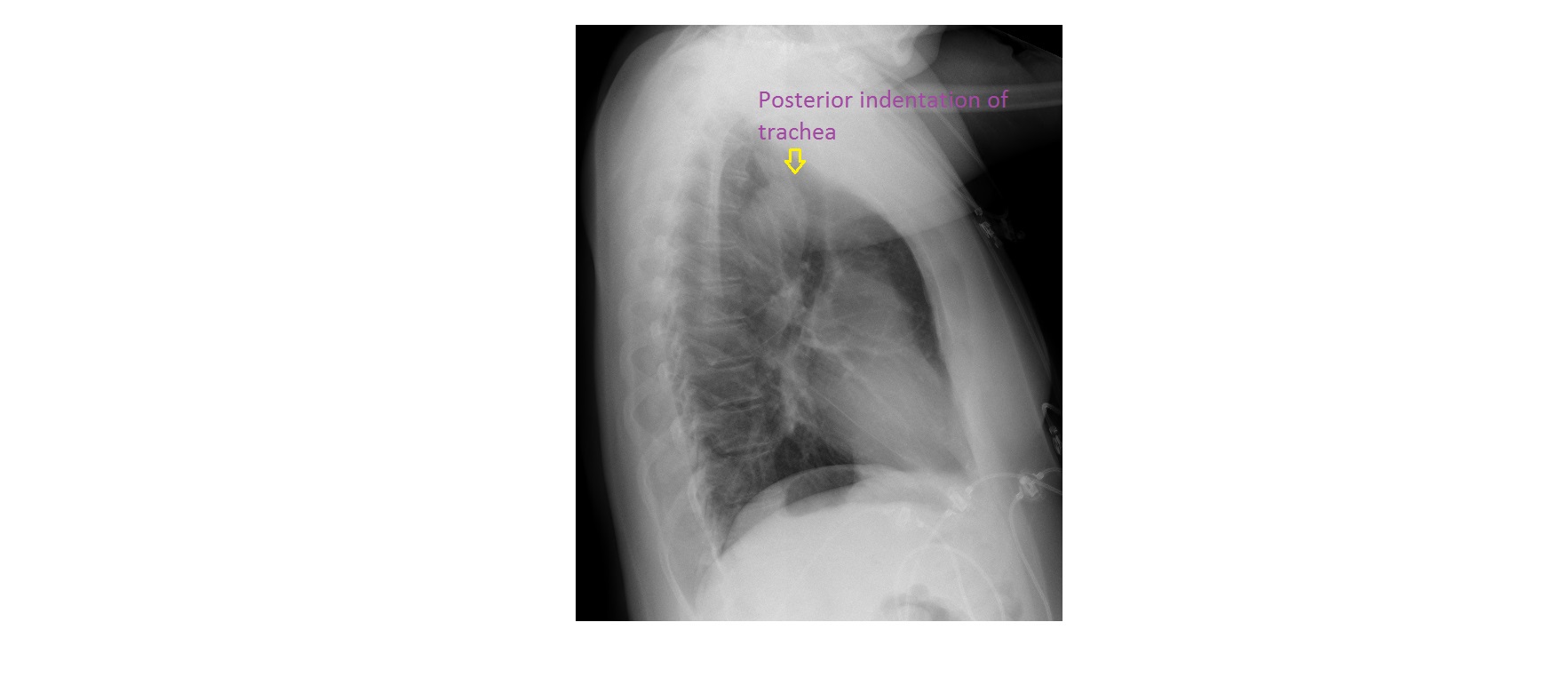
The lateral chest x-ray shows posterior indentation of the trachea.
There is no cardiomegaly and rest of the lung fields are clear.
Echocardiogram confirmed a right sided aortic arch. The retrotracheal bulge seen is likely due to aberrant left subclavian artery.
Incidence of right sided aortic arch is about 0.1%. It can be associated with cardiac abnormality such as VSD. Echocardiogram will help to confirm the diagnosis as well as rule out cardiac abnormality.
Patients with a right sided aortic arch can present to the ED with proximal airway obstruction or dysphagia due to proximal oesophageal constriction.
In the above patient, it was an incidental finding.