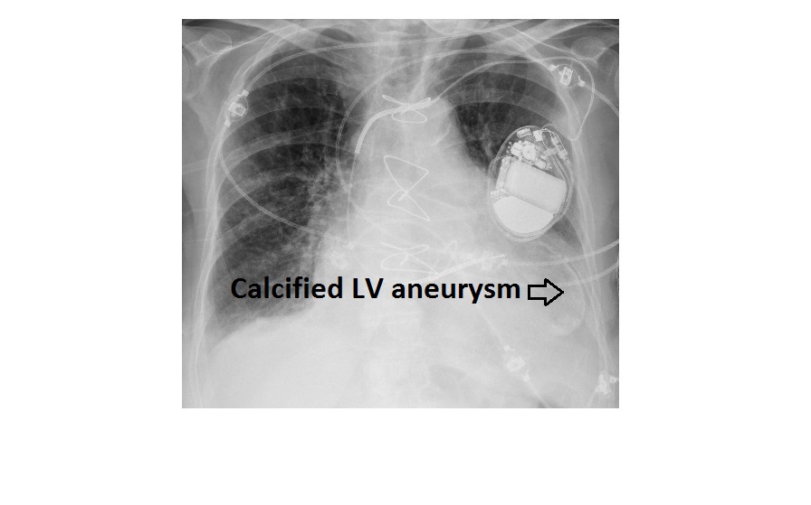
The frontal chest x-ray shows a curvilinear calcification in the area of cardiac apex, in keeping with a calcified LV aneurysm. An echocardiogram confirmed the presence of LV aneurysm.
There is cardiomegaly despite the image being an AP view. There is an implantable cardiac defibrillator and a metal mitral valve. There are sternotomy wires as well as ecg leads.