
The frontal knee x-ray looks normal. The HBL view shows a knee effusion, as noted by the separation of the pre-femoral and supra-patellar fat pads by more than 10 mm.
The HBL view of the knee also reveals a lateral notch sign, as indicated by an abnormally deep indentation of the condylo-patellar sulcus of the lateral femoral condyle. The lateral notch sign is a specific, but not sensitive, sign for ACL (anterior cruciate ligament) injury.
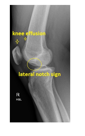
This patient went on to have an MRI of the affected knee, which showed an impaction fracture of the condylo-patellar sulcus of the lateral femoral condyle as well as a complete mid-substance tear of the ACL.
Reference: Grainger and Allison’s Diagnostic Radiology, A Textbook of Medical Imaging, 6th edition.