
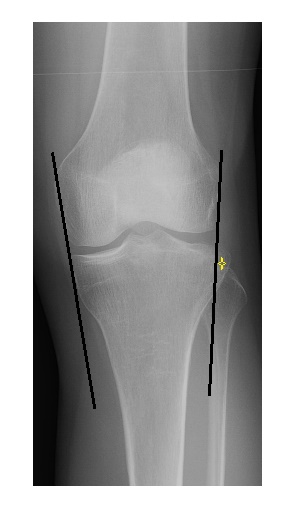
The frontal knee x-ray shows an irregular contour of the lateral tibial plateau.
The HBL view shows lipohaemarthrosis.
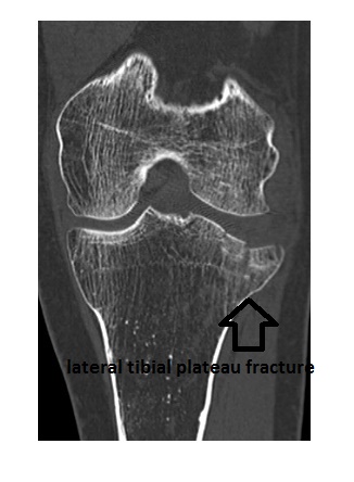
A CT scan showed a compression fracture of the lateral tibial plateau with 5mm depression of the articular surface.
Tibial plateau fractures are difficult to diagnose on plain x-rays. An indirect sign of intra-articular knee fracture is the presence of lipohaemarthrosis (fat-fluid interface) on the HBL view.
Other useful clues are:
- Increased density over the tibial plateau due to bone compression.
- A perpendicular line drawn at the most lateral margin of the femur should not have more than 5mm of the adjacent margin of the tibia beyond it.
The above patient underwent operative intervention.
Reference: Accident and Emergency Radiology: A Survival Guide by Raby et al, 2nd edition.
Excellent sharing. Thank you so much.