
The lateral ankle x-rays show the obliteration of the pre-Achilles fat pad (i.e. Kager’s triangle). With a high clinical suspicion for a tendoachilles tear, the two patients underwent an ultrasound scan of the heel and in both cases, a complete Achilles tendon rupture at the myotendinous junction could be observed.
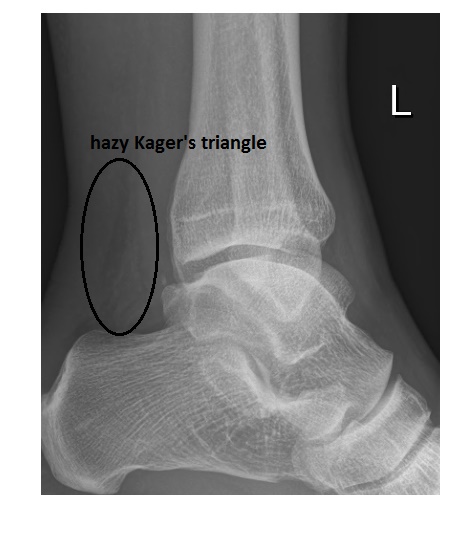
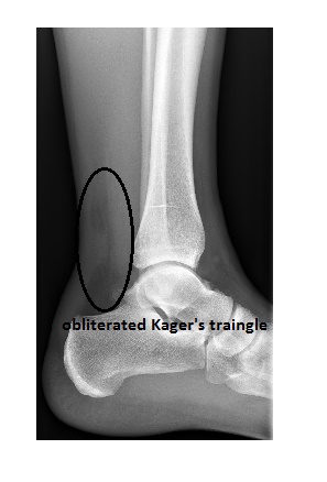
Kager’s triangle is a pre-Achilles fat pad visible in the form of a well defined triangle posterior to the tibia on a lateral ankle x-ray. Its anterior border is formed by the FHL (flexor halluces longus tendon), and its posterior border by the Achilles tendon; the calcaneus forms the inferior border.
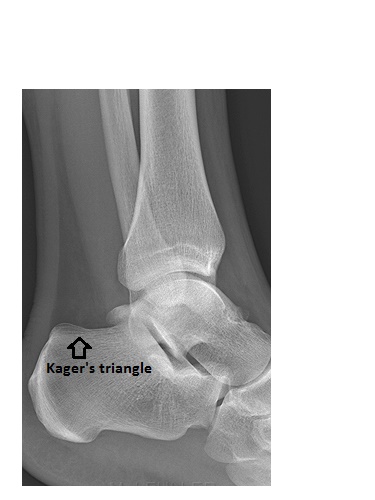
The triangle is distorted in various conditions around the ankle, including Achilles rupture.
Reference here.