
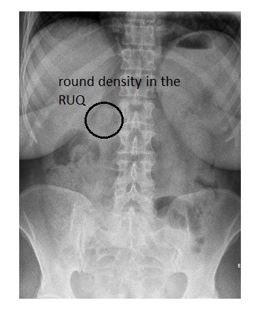
The erect abdominal x-ray does not show small or large bowel dilatation. There is no air fluid level seen. There is a large round density seen in the right upper quadrant, likely a gall stone.
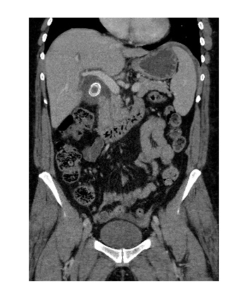
The patient subsequently underwent a CT scan which showed a large 2.5 cm gall stone at the neck of the gall bladder causing distension of the gall bladder with wall thickening and oedema.
The patient was admitted under the specialist and underwent cholecystectomy.
Less than 10% of gall stones are radiopaque on plain abdominal x-rays. Ultrasound is the most accurate investigation for the diagnosis of gall stones.
Reference: Grainger & Allison’s Diagnostic Radiology, 6th Edition.