
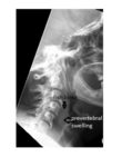
The lateral neck x-ray shows a curvilinear density in front of the C4 vertebra which, considering the history of ingestion of fish, is likely a fish bone. There is also an associated swelling of the pre-vertebral soft tissue.
 The patient underwent oesophagoscopy for removal of the fish bone.
A lateral neck x-ray is not a sensitive test to identify the presence of a fish bone in the oesophagus, and the level of visibility depends on the radiodensity of the fish bone itself. As such, a normal lateral neck x-ray does not rule out the absence of fish bone in the upper digestive tract.
Indirect signs include a widened pre-vertebral space (which is normally smaller than the width of the adjacent vertebra) and the presence of gas/air-fluid level in the pre-vertebral space.