
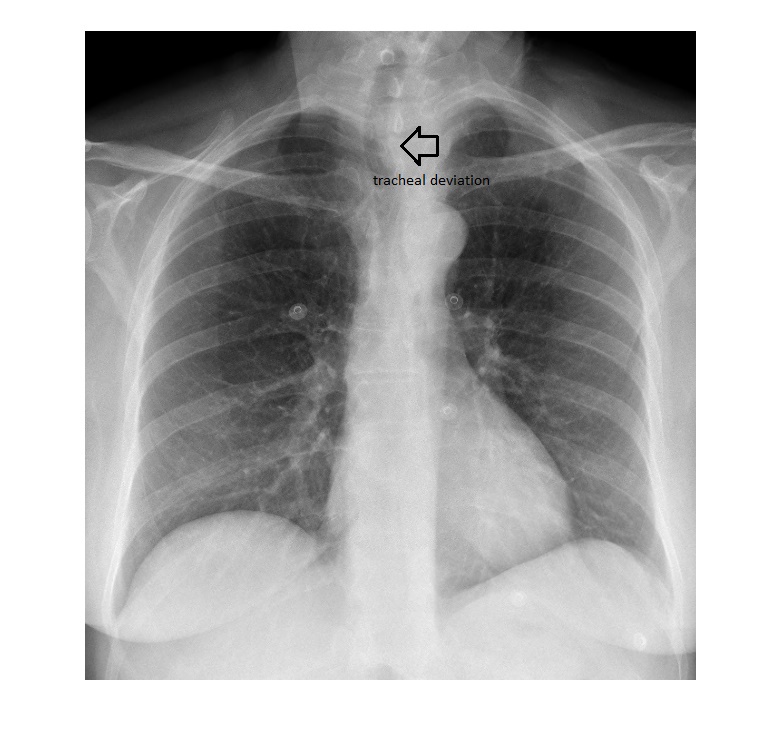
The chest x-ray shows clear lung fields and a normal cardiac size. The x-ray is slightly rotated; however, the trachea appears to be deviated to the right. In addition, a left para-tracheal haziness is also observed.
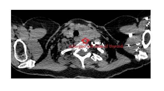
 The patient underwent a CT scan of the chest, which showed an enlarged left lobe of the thyroid, measuring approx. 6×3 cm.
While interpreting a chest x-ray, review areas should be carefully scrutinised. These include the lung apices, hilum, and the retro-cardiac and sub-diaphragmatic areas.
The patient underwent for X-ray and CT scan which means his money is spent on two types of scans even in the presence of MRI technology. We believe MRI could have shown a clear result in a single scan and the patient would not need any further scanning and also be helpful in diagnosing.
Hi Dr. Pegram,
Thank you for your feedback. Kindly note that I am an Emergency Physician. The chest x-ray was done as a part of emergency evaluation of chest pain in a 64 year old. CT scan of the chest was done as there was a concern regarding pulmonary embolism.
Thank you.
Dr. Shenoy