
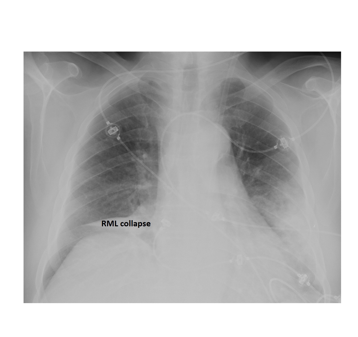
This chest x-ray is from a 70 year old who has presented with dyspnoea and fever. What can you see?

The chest x-ray shows right middle lobe collapse and patchy air space opacities involving the left lower and mid zones.
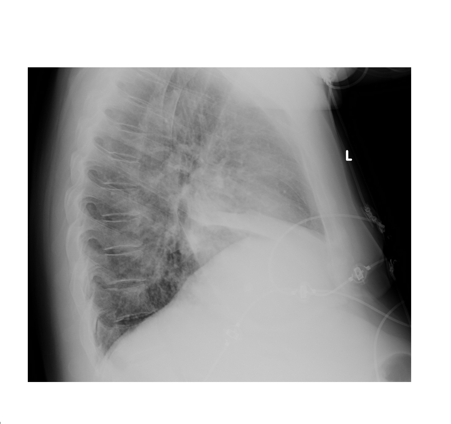
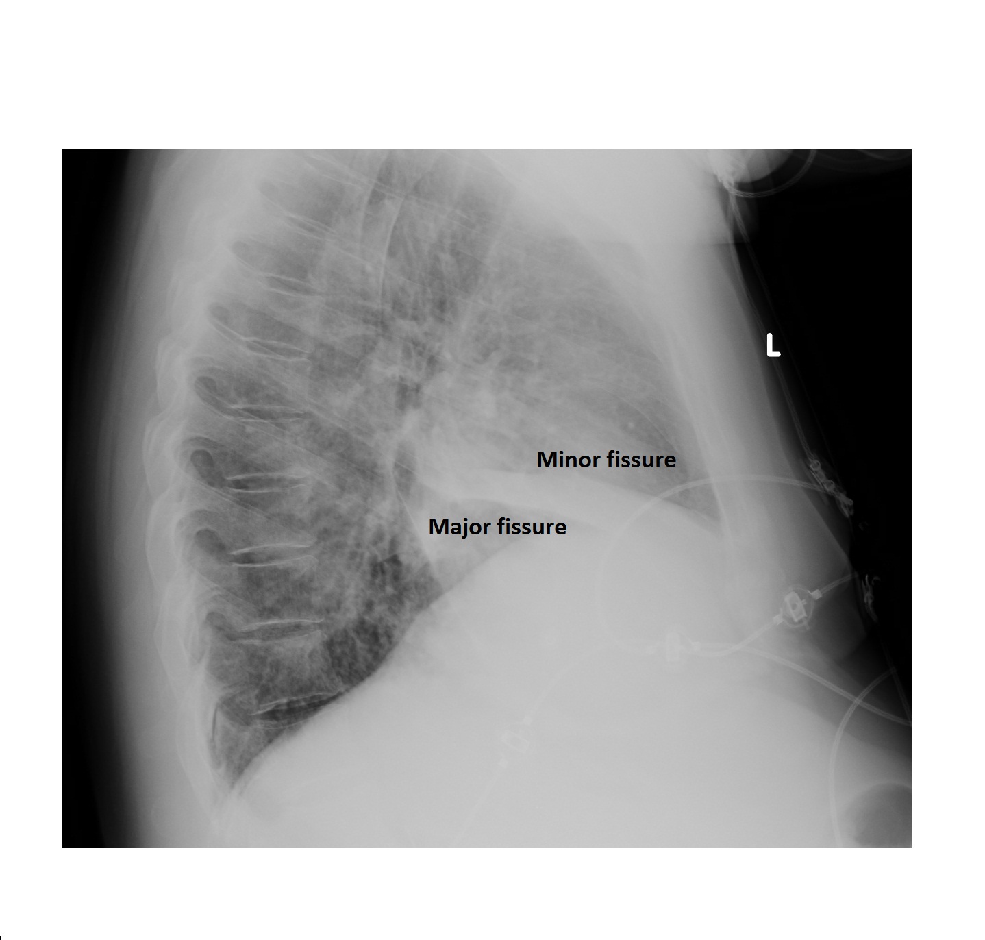
Right middle lobe collapse is visible on the x-ray as a density silhouetting the right heart border. The associated volume loss has resulted in movement of the minor and the major fissures as seen on the lateral view, overlying the heart shadow.
Three causes of indistinct right heart border on chest x-ray are:
- Right middle lobe collapse (volume loss; minor and major fissures pulled towards the dense opacity).
- Right middle lobe consolidation (see imaging case of the week 24; no volume loss, may have air bronchogram and the fissures stay in place).
- Pectus excavatum (the depressed sternum displaces the heart to the left; lateral chest x-ray confirms the sternal deformity).