The following frontal chest x-ray is from an 80 year old with lethargy. Patient is noted to have hypercalcaemia on blood tests. What can be seen on the chest x-ray?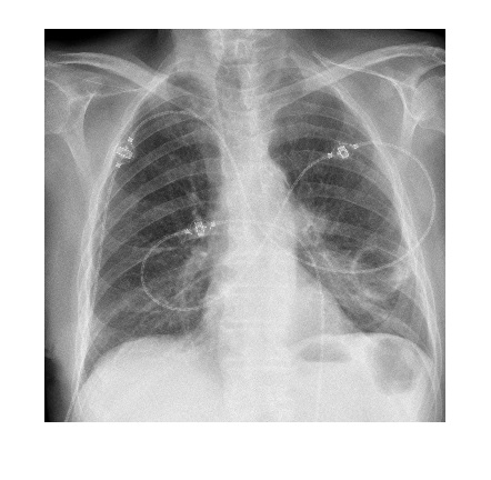 Answer will be posted in a few days.

The following frontal chest x-ray is from an 80 year old with lethargy. Patient is noted to have hypercalcaemia on blood tests. What can be seen on the chest x-ray?Answer will be posted in a few days.
Some peri-bronchial thickening bilaterally, ?lymphadenopathy to Left hilum
well defined calcified lesion to Left Lobe ?Granulomatous mass from TB/Sarcoidosis
with hypercalcaemia ?lymphoma/malignancy