
The frontal chest x-ray shows features of PE (pulmonary embolism):
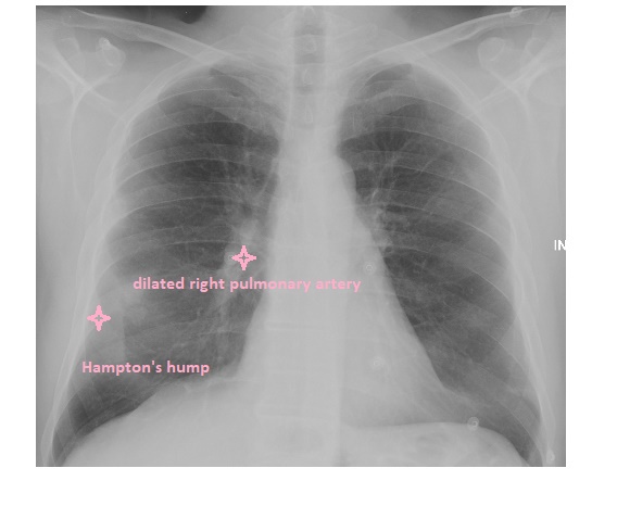
There is a peripheral pleural-based opacity in the right mid zone – this is Hampton’s Hump, which represents pulmonary infarction.
There is distension of the right pulmonary artery with attenuation of the vessel distally and this known as the knuckle or sausage sign.
In addition, the left lower-lobe pulmonary artery shows an abrupt cut off.
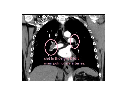
The patient had a CTPA subsequently which showed bilateral central pulmonary emboli.
In a patient suspected of having a PE, a chest x-ray is undertaken to rule out other mimics such as a pneumothorax or pneumonia, as opposed to diagnosing the PE.
Reference: Grainger & Allison’s Diagnostic Radiology, a Textbook of Medical Imaging.