
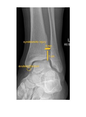
The left ankle mortise view shows syndesmotic injury. The tibiofibular clear space is wide, measuring about 9 mm. The tibiofibular clear space, when measured 1 cm above the tibial plafond should be less than 6 mm in width.
Left leg x-ray did not reveal a proximal fibular fracture (Maisonneuve injury). The patient had a syndesmotic repair by the orthopedic surgeon.
Isolated syndesmotic injuries can be difficult to diagnose radiologically. Comparative stress weight bearing x-rays can be helpful. If there is a high index of suspicion, advanced imaging (MRI) can help.
Reference: www.orthobullets.com, http://www.imagingpathways.health.wa.gov.au/index.php/imaging-pathways/musculoskeletal-trauma/bone-and-joint-trauma/ankle-injury#pathway