
A 24 year old man has been punched in the face and presents with pain and bruising around his left eye. He is intoxicated. The radiographer has been able to obtain only one view (OM view) due to patient non-cooperation. What does it show?

The facial xray shows tear drop sign on the left which suggests the presence of a blow out fracture. The tear drop represents herniated orbital floor contents. An orbital blow out fracture is associated with fracture of one of the walls of orbit with the rim remaining intact.

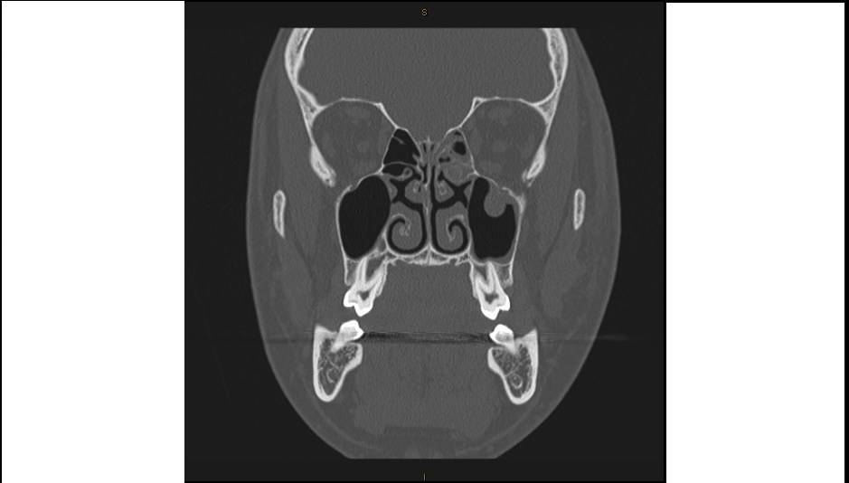
Here is the CT scan of the same patient which shows the anatomy more clearly.
The other indirect radiological feature of inferior orbital wall fracture is the presence of fluid in the maxillary antrum which represents blood in the context of trauma. There may be a black eyebrow sign due to air from the antrum entering the orbital cavity which is visible on plain radiographs just below the superior orbital margin.
Clinical features of orbital blow out fractures are: enophthalmos, parasthesia over cheek and upper lip on the ipsilateral side, vertical diplopia on upgaze due to inferior rectus tethering and crepitus around eye.
These patients need referral to maxillofacial surgeons. Presence of orbital emphysema should alert you to the possibility of orbital compartment syndrome which is an ophthalmological emergency.
[/peekaboo_content]