
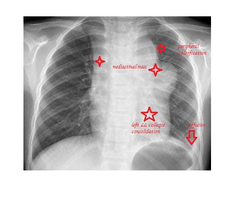
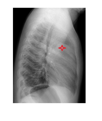
The chest x-rays show an anterior mediastinal mass. The left hilar area shows lobulated appearance with irregular edges and a peripheral rim of calcification. There is volume loss in the left hemithorax. There are additional findings of left lower lobe consolidation with collapse & a left pleural effusion.
The causes of anterior mediastinal mass are – 5 T’s
- Thymoma
- Teratoma
- Thyroid mass
- Terrible lymphoma
- Thoracic aortic aneurysm
Reference: Fundamentals of Diagnostic Radiology, Brant & Helms.