
The following knee x-rays are from a 40 year old man who is complaining of mild knee pain after a trivial fall. He does not have any point tenderness on the knee and is able to fully weight bear on the affected leg. What can you see ?
click to enlarge
click to enlarge
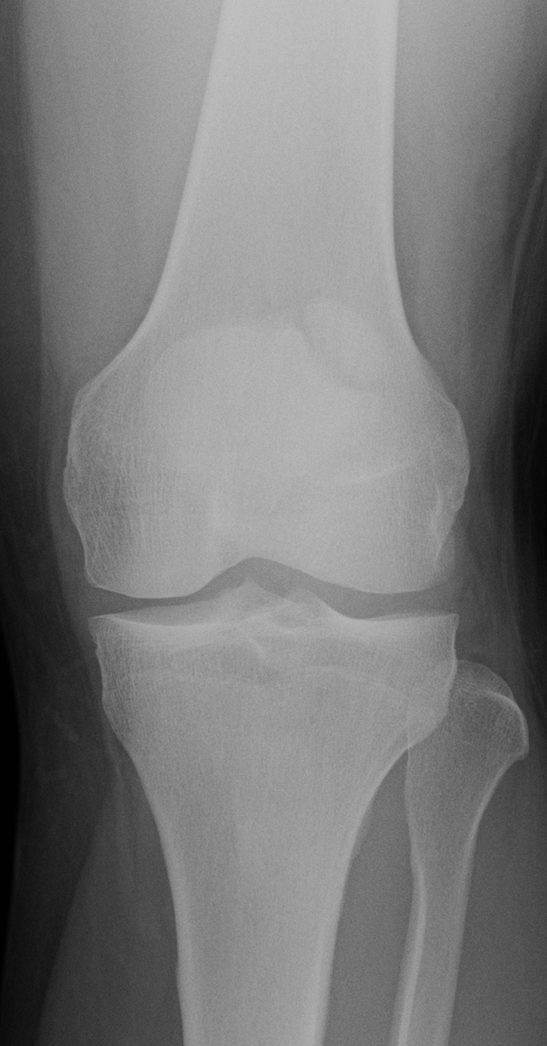
The knee x-rays show no fracture. However, there is an abnormality in the AP view with a well corticated segment of patella situated superolaterally.
This is an incidental finding of bipartite patella which is due to the presence of an accessory ossification centre at the superolateral pole.
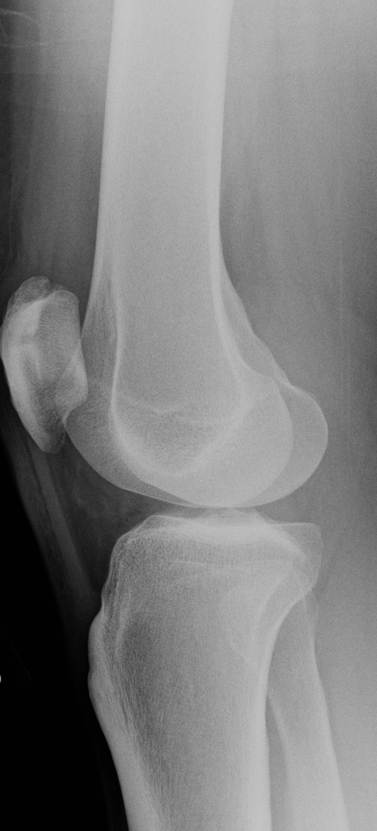
The lateral view does not show a fat fluid sign/lipohaemarthrosis.
Bipartite patella is commonly mistaken for a fractured patella. A fractured patella has the following features:
- It is associated with haemarthrosis of the knee
- Fracture fragments will have an irregular outline
Correlating x-ray findings with clinical examination is important; there will be point tenderness in a fractured patella and the patient may be unable to perform a straight leg raise.

click to enlarge