
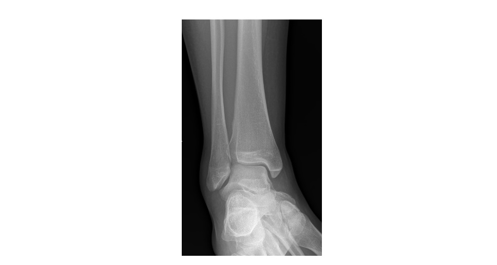
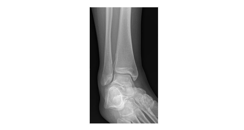
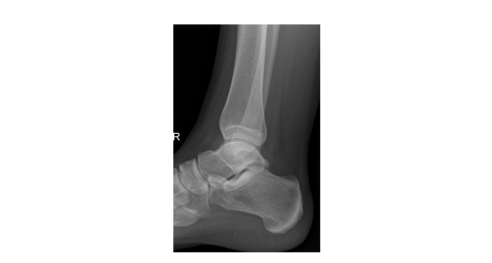
The following ankle x-rays are from a 22 year old woman who twisted her ankle when falling down the stairs. Her ankle is swollen and she is unable to weight bear. Can you spot the two injuries she has ?
click to enlarge
click to enlarge
click to enlarge
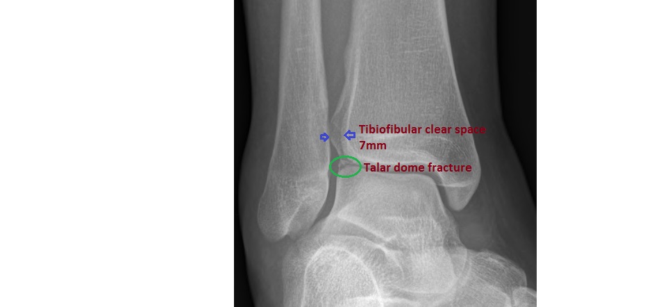
The ankle x-rays show:
- Talar dome fracture – there is osteochondral fracture of the lateral corner of the talar dome.
- Syndesmotic injury – there is widening of the tibio-fibular clear space. The width of the tibio-fibular clear space when measured 1cm above the ankle joint should be less than 5mm in both AP and mortise views. In the above x-rays, however, it measures almost 7mm, indicating diastasis.
click to enlarge
Isolated syndesmotic injuries are sometimes referred to as ‘high ankle sprains’. It is important to recognise these injuries in the ED as they need referral to an orthopaedician for a syndesmotic screw fixation.
(Reference: http://www.wheelessonline.com/ortho/objective_diagnosis_of_syndesmotic_injury)
Thanks to Dr. John Larkin for the images.
[/peekaboo_content]
great case … subtle but significant injuries