
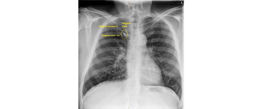
This chest x-ray is from a 55 year old man who presented to the ED with dyspnoea. Largely, it looks normal. However, there is a subtle anatomical variant visible on the x-ray. Can you spot it?

click to enlarge
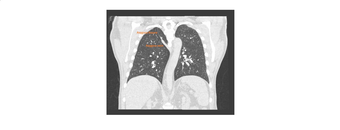
There is an azygous lobe, a developmental anomaly seen in about 1% of the general population. It is a small accessory lobe found on the right lung apical area.
The azygous fissure is visible as a fine convex line which separates the lobe from the rest of the upper lobe. The thickened lower part of the fissure contains the azygous vein.
click to enlarge
click to enlarge
It is of no clinical significance. However, the end-on appearance of the azygous vein can sometimes be confused for a pulmonary nodule.
[/peekaboo_content]