
The following knee x-rays are from a 33 year old patient who landed on his feet after a fall from a height. His knee is swollen and he is unable to weight-bear. What can you see?

click to enlarge
click to enlarge
The AP view of the knee shows a small fracture fragment just outer to the lateral tibial condyle, suggestive of Segond’s fracture.There is also an ill-defined bone fragment in the medial tibio-fibular joint space.
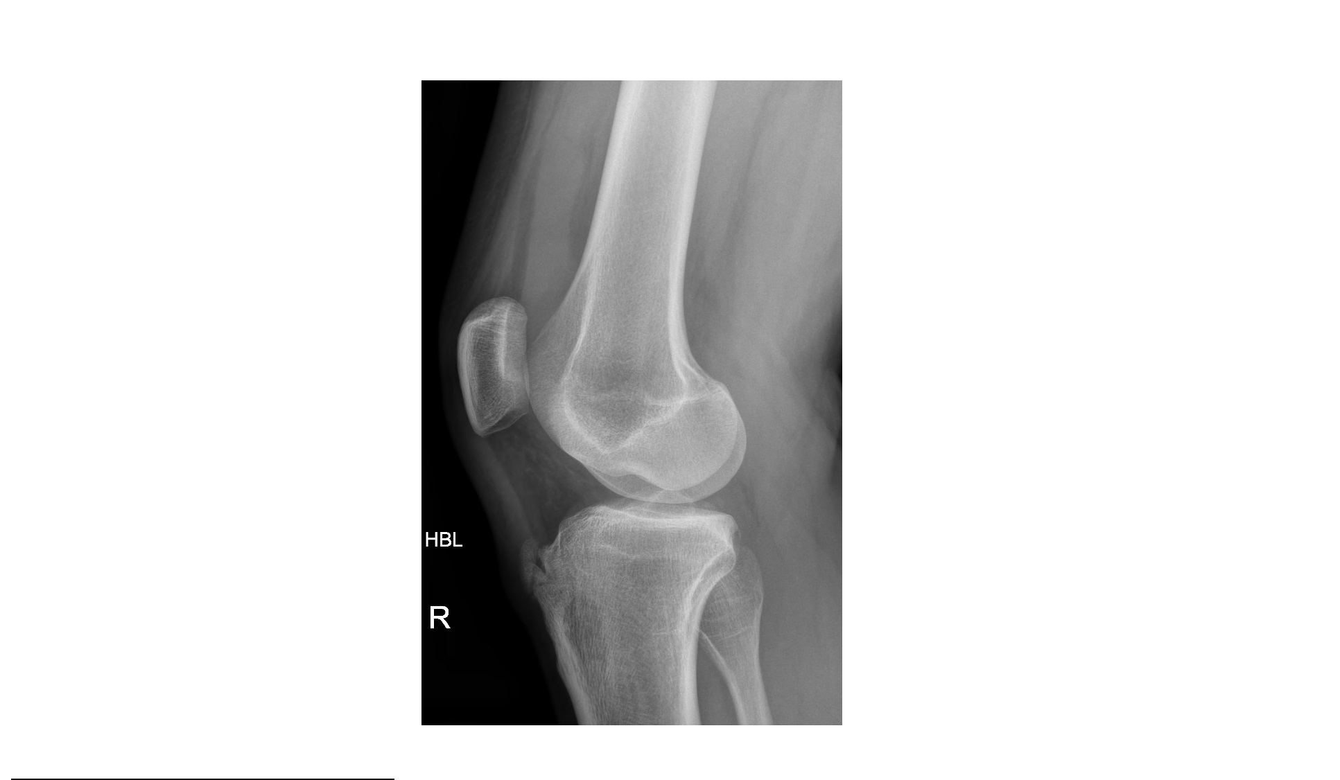
The lateral view shows moderate effusion (lipohaemarthrosis).There is focal indentation of the lateral femoral condyle, which is indicative of a compression type fracture.

click to enlarge

click to enlarge
The above x-rays may show only small fractures but they signify considerable internal derangement of the knee joint. The patient had a completely torn ACL and tear in the posterior horns of the medial and lateral menisci.
[/peekaboo_content]