
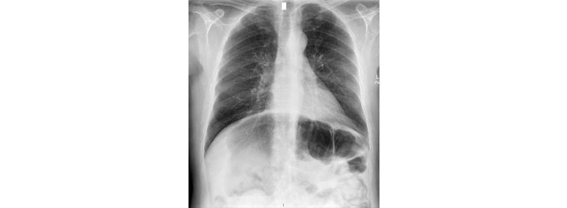
The following abdominal x-rays are from a 65 year old with abdominal distension. There is an obvious coffee bean sign; which part of the large bowel is twisted?

click to enlarge
click to enlarge
There is a grossly dilated, inverted loop of the large bowel with a maximal diameter of up to 12cm. The haustra are clearly seen traversing only part of the bowel circumference.
The affected large bowel is seen pointing to the right upper quadrant. This is sigmoid volvulus. There is no evidence of perforation on the xrays.
The differential for this case would be caecal volvulus – but the caecum is in normal position and the calibre of the caecum and ascending colon is normal.

click to enlarge
Sometimes, it might be difficult to differentiate between sigmoid and caecal volvulus on plain x-rays and a CT scan may be the investigation of choice.
Sigmoid volvulus, in general, affects the elderly who have a history of severe constipation and patients with severe psychiatric illness or a neurological condition.
In contrast, caecal volvulus is more common in younger age groups and in those who have had previous abdominal surgeries.
[/peekaboo_content]