
A 5 year old presents with FOOSH injury and a swollen elbow. His elbow x-rays are as follows. What do you notice?

click to enlarge

click to enlarge
The AP view shows no obvious fracture and the radiocapitellar line is intact.
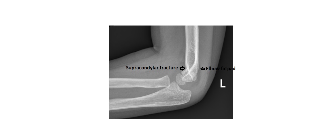
However, the lateral view shows the ‘sail sign’; the anterior humeral line has the entire capitulum lying in front of it. This should raise the possibility of a supracondylar fracture. If you look carefully, there is a supracondylar fracture with volar angulation of the distal humerus.
click to enlarge
More details on fat pad sign and anterior humeral lines can be found at:
http://www.emergucate.com/2012/10/08/imaging-case-of-the-week-16/
[/peekaboo_content]
Effusion + anterior humeral line not passing through middle third capitulum = supracondylar fracture.
However I believe this one of the very uncommon “flexion type” supracondylar fractures where the distal fragment is displaced anteriorly instead of the usual posterior displacement. Usually with supracondylar fractures, they occur in extension where they posterior displacement of the distal fragment means the anterior humeral line passes through the anterior third of the capitulum (instead of the middle third) or it misses the capitulum entirely passing anterior to it. However in this case the anterior humeral line passes posterior to the capitulum. This apparently only occurs in 2% of cases.
Information re classification of supracondylar fractures
http://www.rch.org.au/clinicalguide/guideline_index/fractures/Supracondylar_fracture_of_the_humerus_Emergency_Department/#Classification