
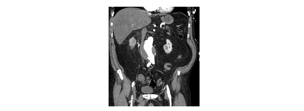
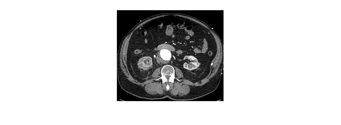
The following CT scan images are from a 74 year old man who has presented with acute onset of right loin to groin pain. What do you observe?
click to enlarge
click to enlarge
The CT images show 2 entirely different co-existing pathologies and both of these can present with acute loin to groin pain.
There is a 6.5 cm infrarenal AAA with mural thrombus. There is no evidence of rupture.
There is also a 7 mm obstructing calculus at the right proximal ureter.
One should always suspect a leaking/ruptured AAA in any elderly patient with new onset of renal colic symptoms.
This patient, who was not unstable, underwent a CT scan with a high index of suspicion for a leaking AAA when he presented with right loin to groin pain. The immediate cause of his symptom turned out to be ureteric calculus. He underwent emergent repair of his aortic aneurysm and had a right ureteric stent placed simultaneously.
[/peekaboo_content]