
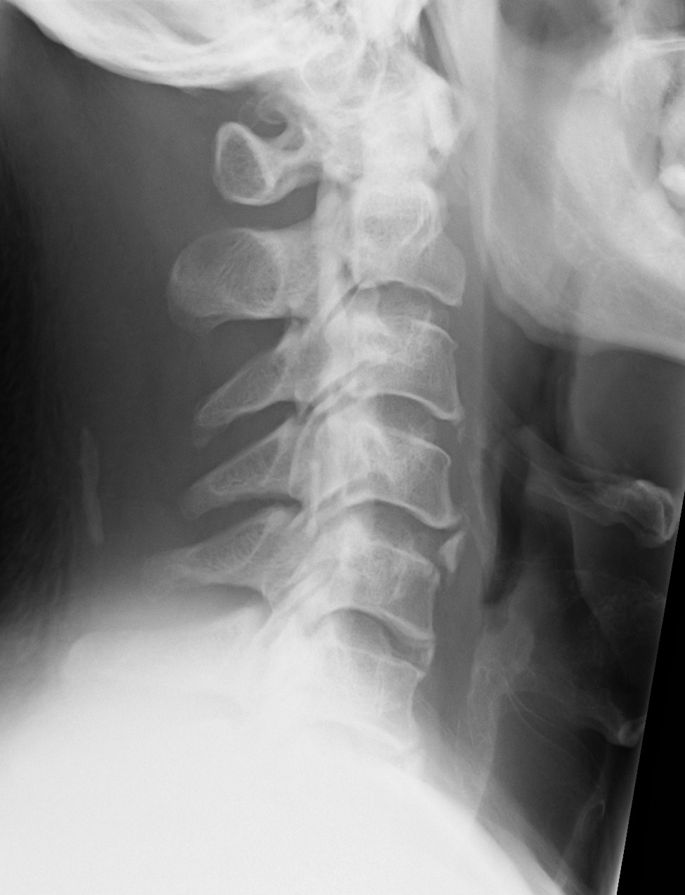
The following lateral c-spine view is from a 54 year old diabetic man with non-traumatic neck pain. What can you see in the x-ray?
click to enlarge
The main abnormality is a linear calcified ropy structure, adjacent to the posterior vertebral margin, projected over the spinal canal. This is an ossified posterior longitudinal ligament (OPLL).
Behind the C4/C5 spine, there is a small linear ossific density in the soft tissue of the neck which represents the ossified ligamentum nuchae.
There is also a focal ossification anterior to the C4/C5 disc which is due to ossification of the anterior longitudinal ligament.
The vertebral alignment and vertebral body height are normal.

click to enlarge
OPLL is an incidental finding and is said to be more common in people of Japanese descent. It is largely asymptomatic but can cause symptoms of cervical myelopathy, at which stage it needs referral to an orthopaedic /neurosurgeon.
[/peekaboo_content]