
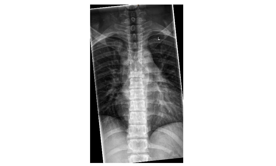
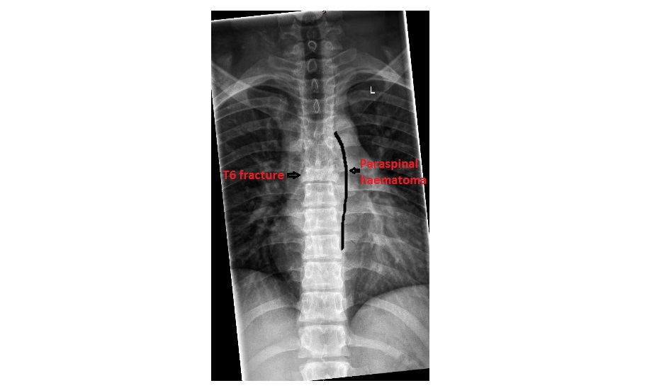
The following thoracic spine x-rays are from a 20 year old male who fell on his feet from a height. There are obvious fractures involving T4 and T6 vertebrae. What soft tissue sign do you notice?
click to enlarge

click to enlarge
The plain x-rays show compression fractures of T4 and T6 vertebrae. The soft tissue abnormality associated is abnormal bulging of the left paraspinal line centred over T6, indicating the presence of a paravertebral haematoma.
click to enlarge
At the thoracic vertebral level, the paraspinal line is formed by the interface between the mediastinal pleura and the paraspinal soft tissues. It is seen as a line that runs parallel to the vertebral bodies from the level of the aortic arch to the T10 vertebra on the left and from T8 to T12 on the right.
Bulging of the paraspinal line indicates posterior mediastinal pathology and in our case, a fractured thoracic vertebra.
References:
[/peekaboo_content]