
A 64 year old man presents with fever, throat pain and difficulty swallowing. He is unable to swallow his own saliva. On examination, he has a soft stridor. You suspect a retropharyngeal abscess with epiglottitis as a differential. What does the lateral neck xray show?

The xray shows swelling of the epiglottis, suggestive of epiglottitis which is an ENT emergency.
Two important radiological signs of epiglottitis are clearly present in this xray.
1. Thumbprinting sign – Swollen epiglottis causes this sign.
2. Vallecula sign – Vallecula is the pre-epiglottic space. It is visible on plain radiographs as a deep air pocket at the level of hyoid anterior to the epiglottis. It is normally parallel to the pharyngotracheal air column. If vallecular sign is present, instead of a deep linear space, a V shaped shallow space is seen.

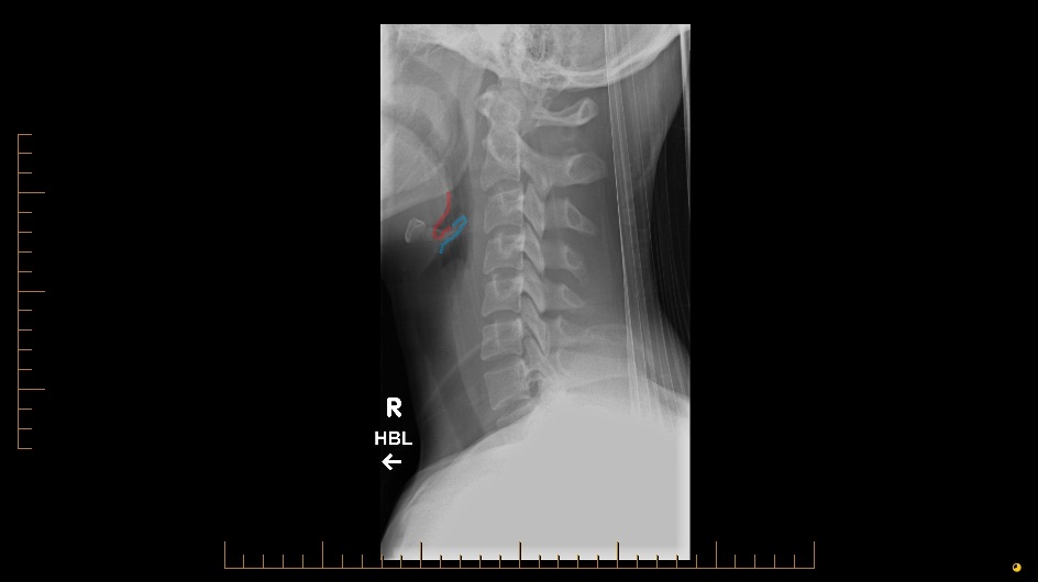
A normal lateral neck xray showing deep vallecular space is presented below for comparison. Red outline demonstrates vallecula and blue outline demonstrates epiglottis.

Note – epiglottitis is largely a clinical diagnosis in ED and lateral neck xray is not a routine investigation, especially in children. Patients should never be positioned supine for the xray as this may close off the airway.
[/peekaboo_content]