
The following chest x-rays are from a 12 year old with a history of cough, fever and dyspnoea who has been unwell for about 6 days prior to presentation. The patient has no past medical history. What can you deduce from the x-rays?

click to enlarge
click to enlarge
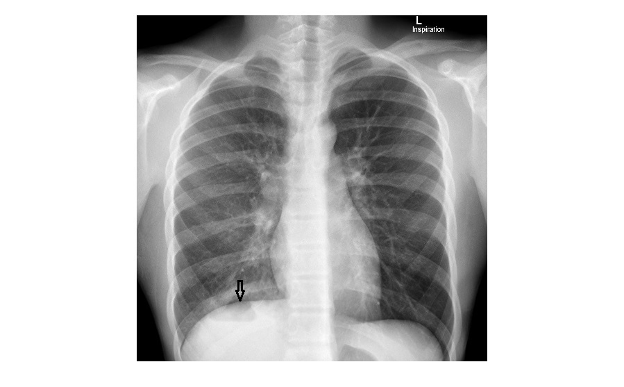
The PA view shows a cavity with fluid level projected over the right dome of the diaphragm with infiltrates in the right basal region.
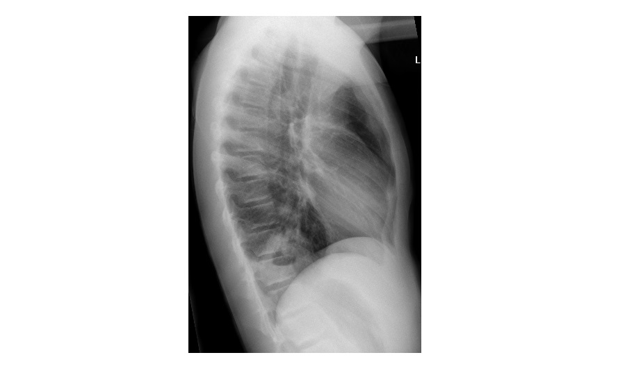
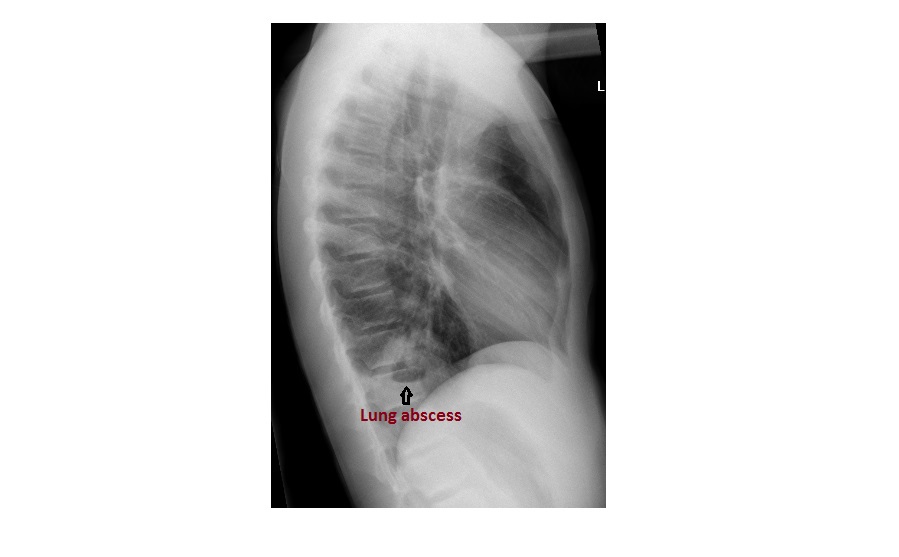
The lateral view clearly shows a thick walled cavity with fluid level in the posterior segment of the right lower lobe.
The findings are consistent with a right lower lobe lung abscess.
click to enlarge
click to enlarge
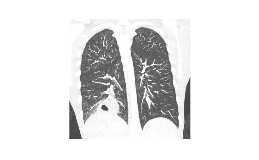
Here are the axial and coronal CT scan slices from the same patient showing a thick walled cavity with fluid level and surrounding consolidation.

click to enlarge
click to enlarge
The patient had high inflammatory markers. Blood culture as well as sputum cultures were negative. He was admitted, commenced on intravenous antibiotics and responded well.
Lung abscesses are less common in children than in adults. Conditions predisposing children to lung abscesses are: aspiration, pneumonia, gastro oesophageal reflux, seizures, immunodeficiency, tracheo-oesophageal fistula and neurological disorders (Nelson Textbook of Paediatrics, 19th ed).
Common pathogens in paediatric lung abscess include streptococcal species, staphylococcus aureus and klebsiella pneumonia (lung abscess in children, paediatric respiratory reviews, Volume 8, Issue 1, March 2007)
Wishing everyone a Happy New Year!
[/peekaboo_content]