
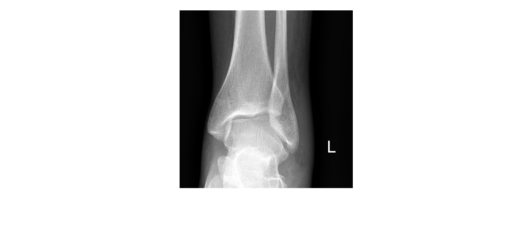
The following ankle x-rays are from a 35 year old patient who sustained a left ankle injury following a fall. She is complaining of pain on the lateral aspect of the ankle and is not weight bearing. What can you observe in the x-rays?
click to enlarge
click to enlarge

click to enlarge
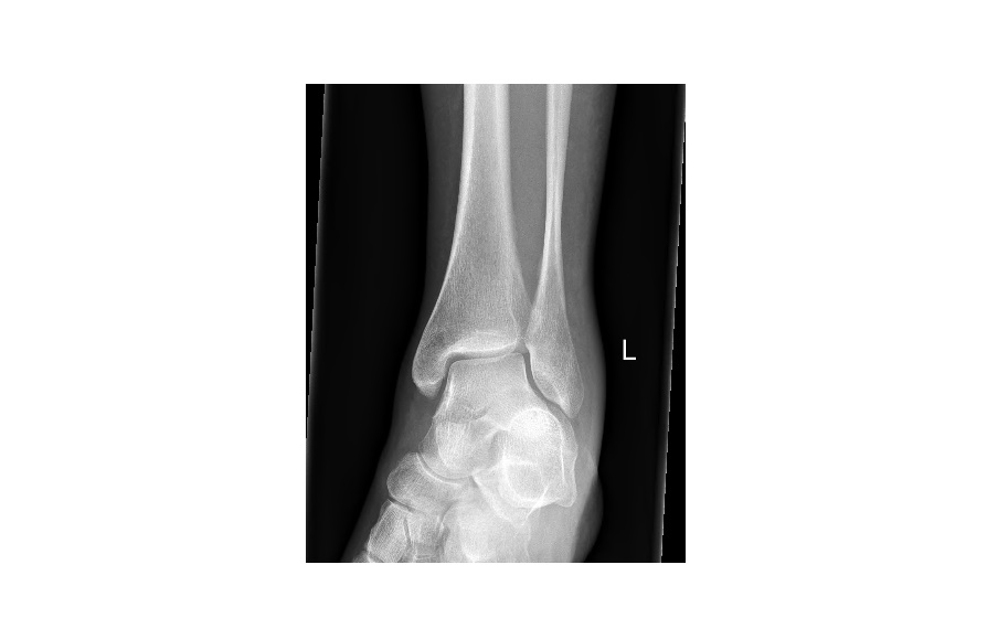
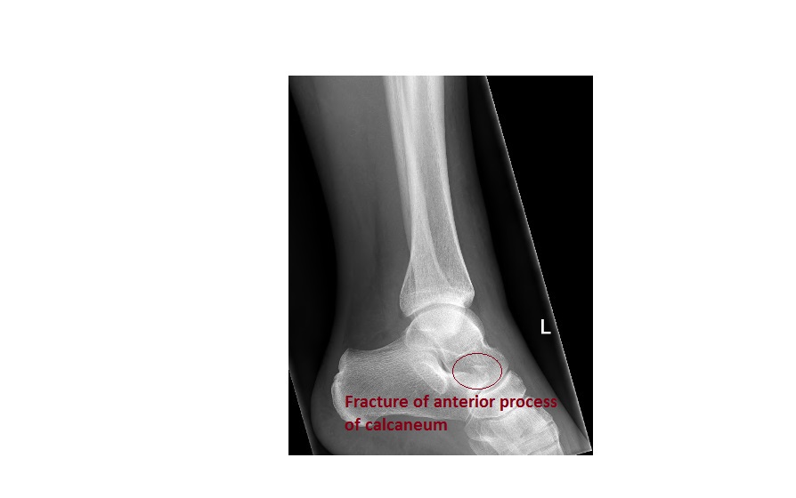
The ankle x-rays show fracture of the anterior process of the calcaneum. There is also soft tissue swelling over the lateral malleolus.
click to enlarge
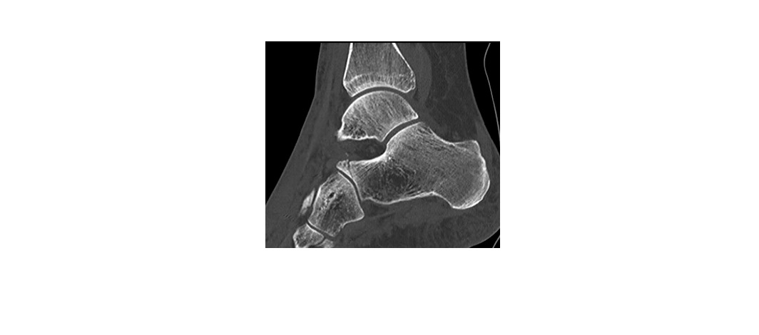
The following CT image is from the same patient.
click to enlarge
The lateral ankle view below, which shows a healing fracture, was taken approximately 8 weeks into the injury.
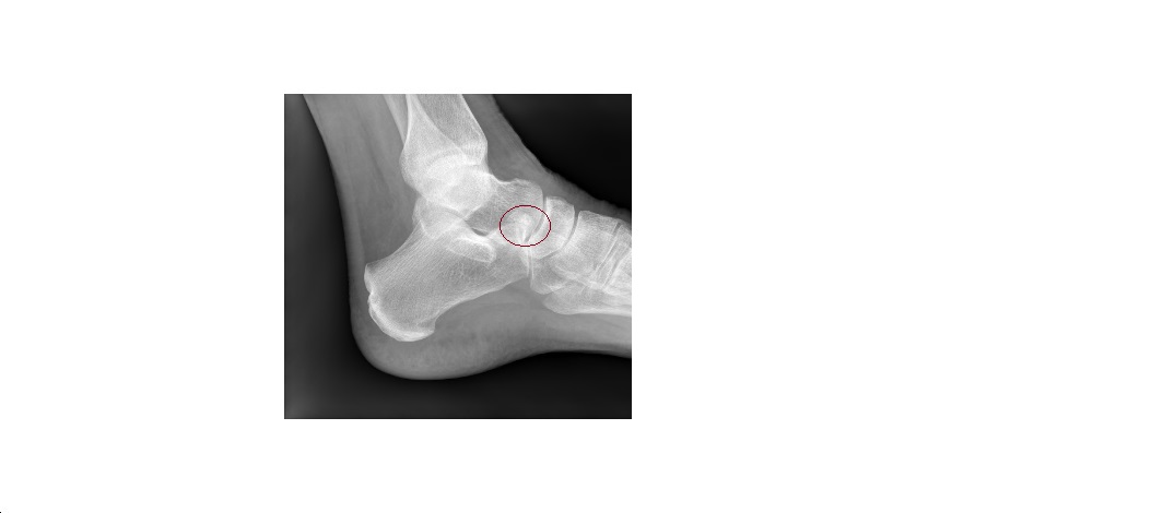
click to enlarge
Fracture of the anterior process of the calcaneum is a missable fracture and is best seen on an oblique foot x-ray.
Mechanism of injury is inversion and plantar flexion of the foot causing avulsion of the bifurcate ligament. The bifurcate ligament connects the anterior process to the cuboid and the navicular.
The patient was treated with cast immobilisation, non-weightbearing and a referral to an orthopaedic specialist for follow up. Indication for ORIF is involvement of >25% of the calcaneocuboid joint.
Our patient was managed conservatively by the specialist.
Reference: http://www.orthobullets.com/trauma/1051/calcaneus-fractures
[/peekaboo_content]