
The following x-ray images are from a 19 year old patient who was involved in a motor vehicle accident. What can you notice on the x-rays?
click to enlarge
click to enlarge
click to enlarge
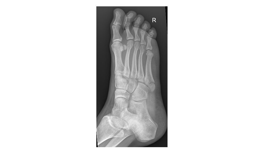
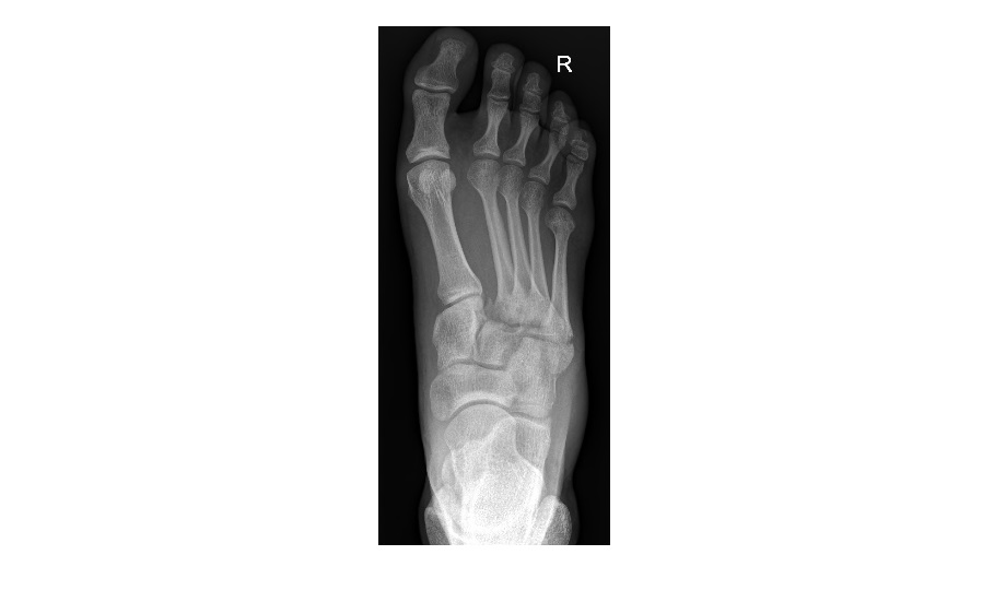
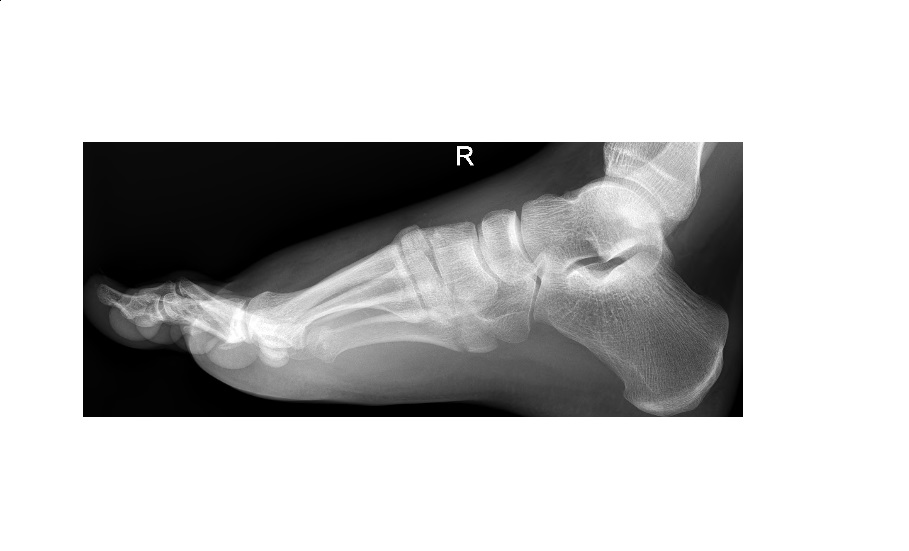
The foot x-rays show a Lisfranc injury with fracture of the bases of the 2nd to 5th metatarsals as well as a lateral displacement of the 2nd, 3rd and 4th metatarsal bases relative to the tarsal bones.
The following 3D CT images are from the same patient showing an abnormally widened gap between the 1st and 2nd metatarsal bases.

click to enlarge

click to enlarge
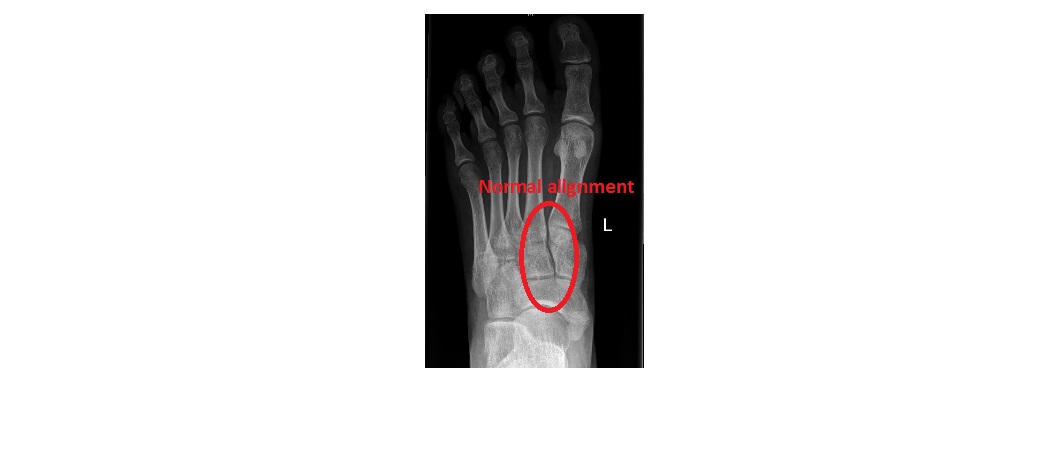
Lisfranc fracture is fracture-dislocation of the tarso-metatarsals. Key to normal alignment is that the medial border of the second metatarsal should always line up with the medial border of the second cuneiform. If the dislocation is minor, it can be easily missed.
click to enlarge
The patient underwent operative reduction and internal fixation.
Reference: Fundamentals of Diagnostic Radiology by Brant and Helms
[/peekaboo_content]