
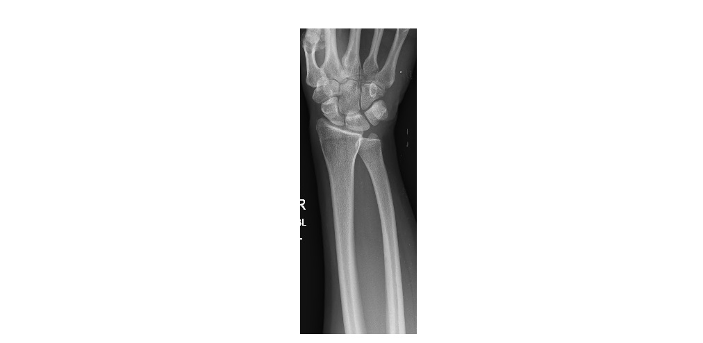
The following wrist and distal forearm x-rays are from a 35 year old woman experiencing wrist pain/swelling following a fall on her outstretched hand. What can be seen?
click to enlarge

click to enlarge
The AP view shows an overlap of the distal radioulnar joint (DRUJ) and the lateral view shows the distal ulna protruding out in a volar direction. This patient has a volar dislocation of the distal radio-ulnar joint.
X-rays of the proximal forearm and elbow did not reveal any proximal injuries.
The dislocation was managed with closed reduction and cast immobilisation in the ED and the patient was referred to the ortho specialist for a follow up.
DRUJ dislocation:
- Can be an isolated injury but can also be associated with a Galeazzi fracture or Essex-Lopresti injuries. As such, it is important to obtain additional entire forearm and elbow x-rays when an isolated DRUJ dislocation is noted on a wrist x-ray.
- Mostly tend to be dorsal; a volar dislocation is less frequent.
- On the frontal radiograph, there will be a widening of the distal radioulnar distance in dorsal dislocations and a superimposition of the radius and ulna in volar dislocations.
- True lateral view will differentiate dorsal from volar dislocations.
- Treated with closed reduction and above elbow plaster with the forearm in supination for dorsal dislocation, and forearm in pronation for volar dislocations.
- Unstable injuries post reduction will need orthopaedic referral for K Wire fixation.
Reference: http://www.med.uottawa.ca/radiology/assets/documents/msk_imaging & http://www.rcsed.ac.uk/fellows/lvanrensburg/classification/wrist/distalradioulnajoint.htm
[/peekaboo_content]
There also appears to be a scaphoid fracture on the AP view.
Hi Belinda,
Thank you for your query.
The wrist film is not a true AP view. As a result, the scaphoid does appear foreshortened. But this patient did not have scaphoid tenderness and subsequent follow up images did not show any scaphoid injury.
Prathibha