
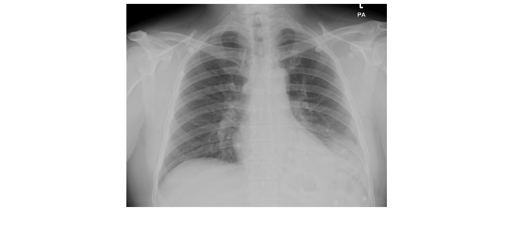
These PA and lateral chest x-rays are from a 40 year old man with a history of fever and dyspnoea. What can you deduce from the x-rays?
click to enlarge

click to enlarge
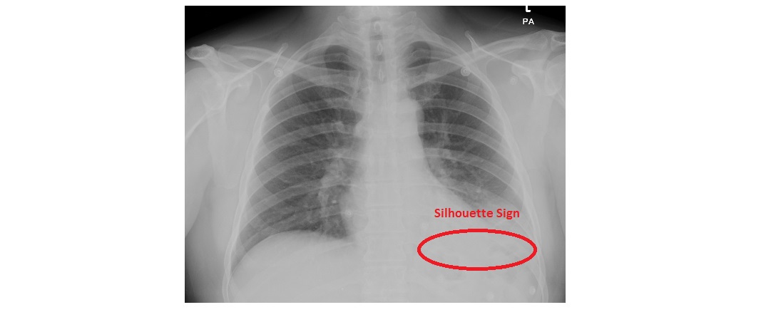
The PA view shows a retro-cardiac and left lower zone opacity, silhouetting the left dome of the diaphragm.
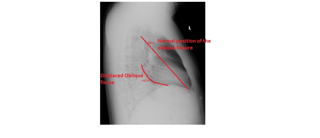
The lateral view shows a triangular opacity behind the heart, with a well-defined superior margin which is the displaced left oblique fissure.The increased density over the lower thoracic vertebra is due to associated consolidation.
click to enlarge
click to enlarge
This patient has left lower lobe collapse consolidation. He was treated with intravenous antibiotics.
Silhouette sign is a loss of contour of the heart or diaphragm, used to localise a parenchymal process. A process involving the medial segment of the right middle lobe obscures the right heart border, a lingular process obscures the left heart border, and a basilar segment lower lobe process obscures the diaphragm.
While dealing with a patient with segmental lobar collapse, causes such as a mucus plug, endobroncheal lesion or a foreign body should be considered.
Any opacity noted on the chest x-ray in the ED needs a follow up film via GP. Bronchoalveolar carcinoma can present as a parenchymal process as opposed to a nodule.
A case of left lower lobe collapse can be found here: http://www.emergucate.com/2012/12/17/imaging-case-of-the-week-26/
Reference: Fundamentals of Diagnostic Radiology, Brant and Helms.
[/peekaboo_content]