
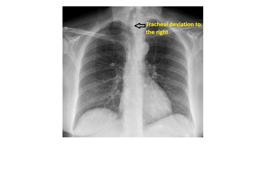
This PA chest x-ray is from a 60 year old female patient who has presented with nonspecific right-sided chest pain. What can you see? (It is an incidental finding)

click to enlarge
The lung fields and heart appear normal. The chest x-ray is mildly rotated.
There is a soft tissue opacity causing smooth indentation of the left lateral trachea with mild luminal narrowing and deviation of the trachea to the right at the thoracic inlet level.
click to enlarge
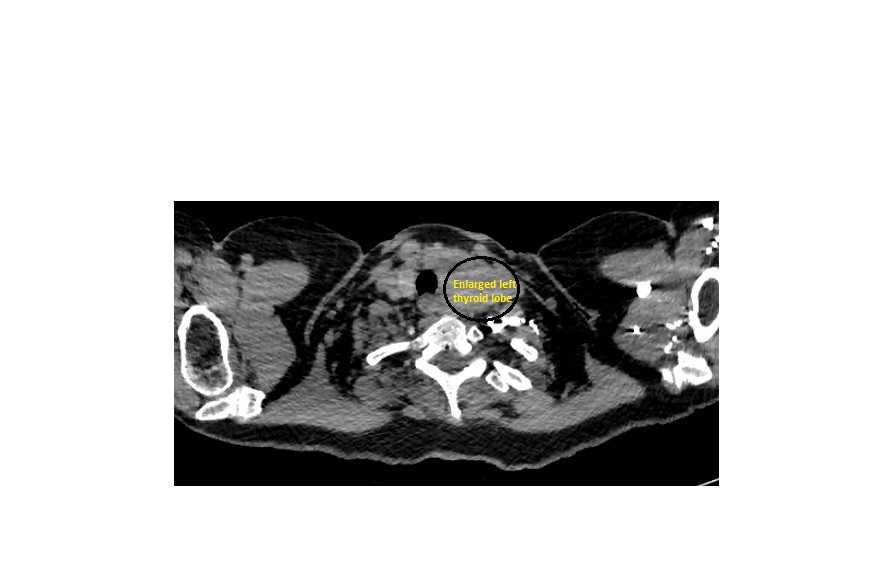
CT scan showed a moderately enlarged left lobe of the thyroid, causing deviation of the trachea; there was no retrosternal extension.

click to enlarge
click to enlarge
Enlarged thyroid (symmetrical or asymmetrical) should be able to be diagnosed clinically as the gland is superficial. However, occasionally a chest x-ray obtained for a different reason may give a clue. In a patient with thyroid enlargement, a plain chest x-ray may show: tracheal deviation, mediastinal thyroid extension (visible as an anterior mediastinal mass), or lung metastasis from thyroid malignancy.
Ultrasound is the initial investigation of choice when a thyroid nodule/enlargement is noted clinically. Ultrasound is also used to obtain FNA from a nodule.
CT is useful when retrosternal extension or extracapsular spread is present.
Reference: http://www.medscape.com/viewarticle/515637_8
[/peekaboo_content]