
The ECG below has been taken from an 89 year old gentleman who has just transferred from the SJA stretcher to an ED bed. He has a history of prostate cancer, IHD and Aortic Stenosis and had an episode of central chest pain with an associated collapse but is now pain free.
Tag Archives: ST changes
ECG of the Week – 8th July – Interpretation
The following ECG has been taken from a 69 year old man who has presented with palpitations and chest pain:
ECG of the Week – 20th May 2020 – Interpretation
ou have reviewed a 45 year old quadriplegic who has a background history of Autonomic dysfunction.
ECG of the Week – 8th April 2019 – Interpretation
Following is an ECG of a 25 years old man with a four day history of left sided chest pain and its 0400 hours in the morning. He’s in a mild discomfort but looks well from the end of the bed and his observation were within normal limits. Describe the abnormalities, differential diagnosis and outline a management plan.
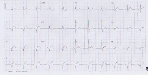
Interpretation:
- Rate: 66
- Rhythm: Sinus rhythm
- Axis: LAD
- Morphology:
- 3mm STE Infr leads
- 2mm convex STE Septolateral leads
- STD V1
- Pathological Q wave inferior leads
- >1mm wide >2mm deep >25% height QRS
- PR elevated aVR
- Intervals: Normal PR and QRS with RBBB pattern
- Summary: ?STEMI – Inferior septolateral ?myo-pericarditis ?coronary vasospasm (given age explore illicit drug use) ?-ve delta wave of WPW (given looks well and obs stable)
Clinical Closure:
- Admitted to cardiology at JHC
- Treated with aspirin / ticagrelor / heparin / analgesia
- CXR normal
- Trop 32000
- Angiogram – normal coronaries with mild focal inferior hypokinesis
- Echo – normal LV, akinesis mid inferior lateral walls, impaired systolic function EF51%, thickened infr-lateral wall (?oedema from myopericarditis), small circumferential pericardial effusion
- Rx of aspirin, Ramipril, metoprolol, ibuprofen
- F/u Echo in 2/12 – resolution of oedema, EF 57%
ECG of the Week – 15th January 2020 – Interpretation
The following ECG is from an 18 year old man who has presented with palpitations and left sided chest pain.
ECG of the Week – 18th September Interpretation
An 83 year old lady presents with palpitations and central chest pain.
ECG of the Week – 24th April 2019 – Interpretation
The following ECG is taken from a 82 year old non-English speaking lady whose family states she woke feeling unwell and with a funny feeling in her left arm:
ECG of the Week – 6th March 2019 – Interpretation
The following ECG is from a 55 year old male who presented to the ED after experiencing on-going left sided chest ache.