
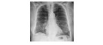
The following chest x-rays are from a 72 year old who has a history of COAD, who has presented with a fever, cough and dyspnoea. What can you infer from the x-rays?
[peekaboo_link name=”Answer”]Answer[/peekaboo_link] [peekaboo_content name=”Answer”]
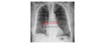
The frontal chest radiograph shows an opacity adjacent to the right heart border causing silhouetting and focal contour abnormality. The right hemidiaphragm appears slightly elevated and there is a degree of hyperinflation of the lung fields.
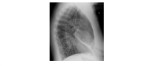
The lateral view shows a linear band-shaped opacity projected over the cardiac shadow. This appearance is classic for complete right middle lobe collapse.
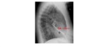
In the presence of infective symptoms, the likely cause is a mucus plug. However, the possibility of an endobronchial lesion should be kept in mind and a CT chest may be needed if there is a failure of resolution of the chest x-ray finding after appropriate treatment.
[/peekaboo_content]