
The chest x-rays are from a 50 year old chronic smoker presenting with chest pain and cough. What can be seen?

[peekaboo_link name=”Answer”]Answer[/peekaboo_link] [peekaboo_content name=”Answer”]
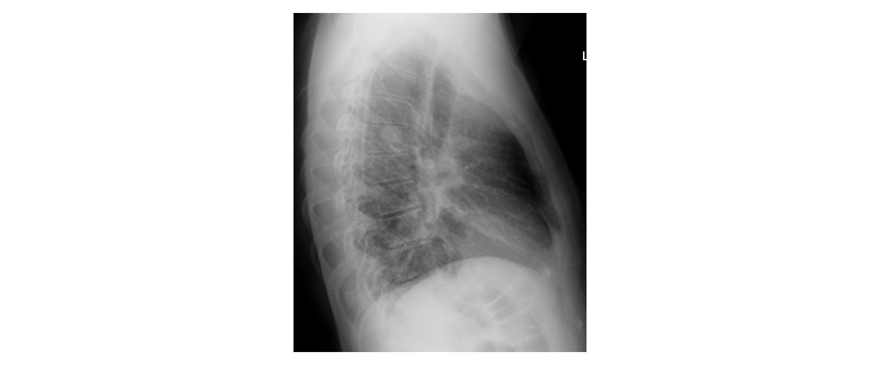
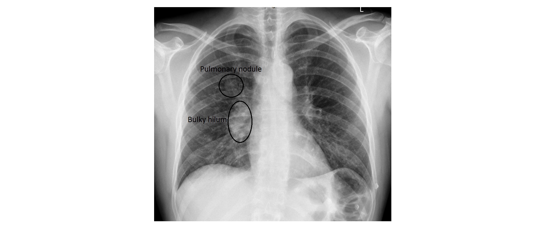
The frontal chest x-ray shows a 2cm nodule in the right mid-zone which is also visible on the lateral view. The right hilum is bulky. The left lung field is clear. These x-ray features are suggestive of a bronchogenic carcinoma.
The patient had a CT scan of his chest for further characterisation of the nodule and it confirmed a primary lung malignancy.
A parenchymal lung opacity, based on the size, is divided into 4 categories:
- Miliary -> less than 2 mm (miliary tuberculosis)
- Micro-nodule -> 2 to 7 mm (acute hypersensitivity pneumonitis)
- Nodule -> 7 to 30 mm (granuloma, metastatic disease)
- Mass -> more than 30mm (bronchogenic carcinoma)
Reference: Fundamentals of Diagnostic Radiology by Brant and Helms, 4th edition
[/peekaboo_content]