The following chest x-ray is from a 40 year old presenting with a fever and cough. What two abnormalities can be seen?
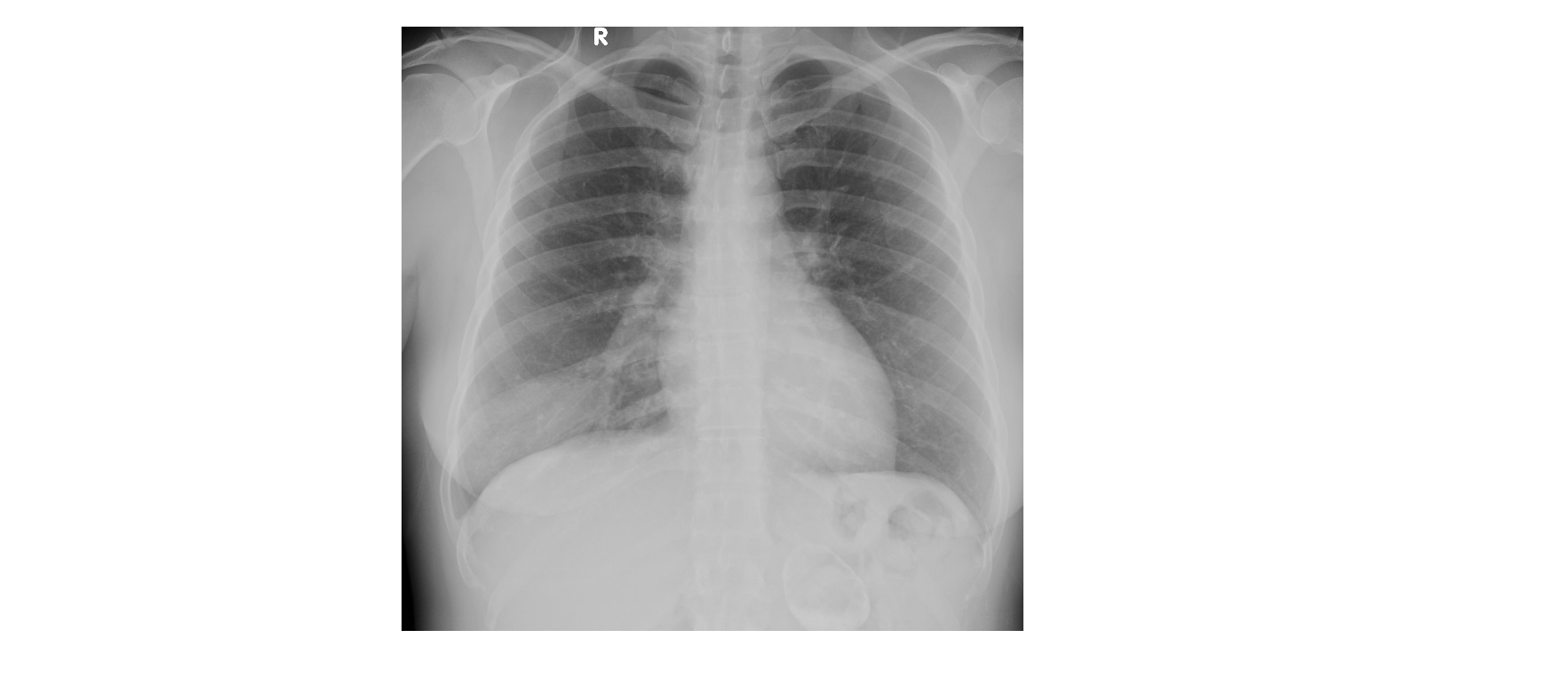
The chest x-ray shows an area of consolidation in the posterior segment of the right lower lobe. With a clinical history of recent onset fever and cough, the x-ray findings are consistent with pneumonia.
The 2nd abnormality is seen as a rounded, peripherally calcified lesion below the left hemi diaphragm (review area). The location indicates either a renal or adrenal lesion.
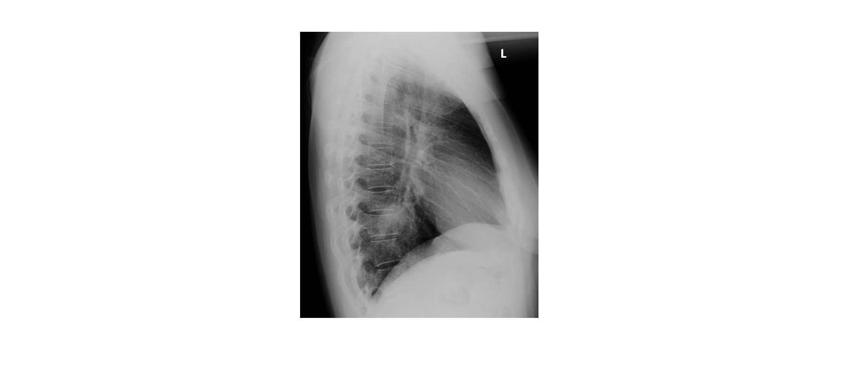
The patient was treated with antibiotics and got better. A repeat chest x-ray six weeks later showed a complete resolution of the consolidation.
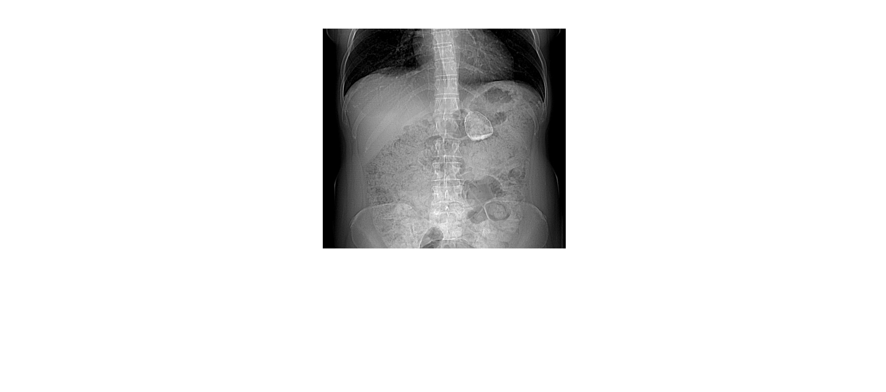
As for the calcified lesion seen on the chest x-ray, she had an elective CT scan of the abdomen which confirmed the lesion to be an old adrenal haemorrhage (I do not have any details on the specifics of adrenal haemorrhage but the patient has remained stable over a period of several years).
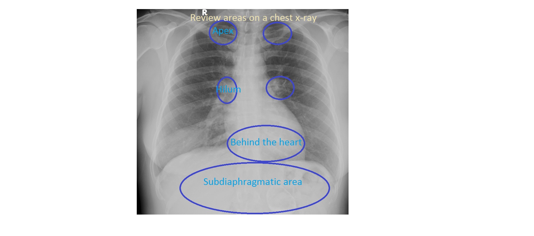
Review areas on chest x-ray are regions where pathology is commonly missed and a careful second look is needed. They are:
- Apices
- Hila
- Behind the heart
- Below the diaphragm