
The chest x-rays show a thick walled cavity in the right middle lobe and an airspace opacity in the left upper lobe with cavity. There is a patchy opacity in the left lingular lobe. There is no pneumothorax or pleural effusion and the cardiomediastinal contour appears normal.
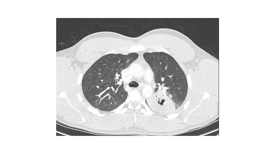
CT scan of the chest showed pulmonary abscess in the left upper lobe and right middle lobe and small basal effusions.

A mnemonic for the causes of cavity in the lung is CAVITY. Details can be found here.
Our patient was treated with long term intravenous anti staphylococcal agent and has been responding to treatment.