
You have just seen a 7 year old girl with a swollen elbow after a fall onto her right elbow. She is in a lot of pain and is very distressed. You obtain an x-ray of the elbow. What can you see?

Click to enlarge

Click to enlarge
There is an obvious large elbow joint effusion. Sail sign is positive and is due to the lifted anterior and posterior fat pads.
Is there a supracondylar fracture? No. The anterior humeral line is passing through the mid capitellum and there is no break in the distal humeral cortex that can be seen.
What about radiocapitellar line? Although there is an alignment in the AP view, there does not seem to be an alignment on the lateral view. There is a radial head dislocation.
A forearm x-ray would be very useful to exclude forearm bone fractures (especially ulnar).
There are 4 essential things to look for in any paediatric elbow x-ray:
- Look for fat pads. There are two-anterior and posterior. Visible anterior fat pad is normal. However, elevated anterior fat pad or any posterior fat pad (either just visible or elevated) is abnormal and indicates associated haemarthrosis. Commonly due to occult supracondylar fracture or radial head/neck fracture in children. Fat pad sign is valid only in a true lateral view with elbow in 90 degree flexion.
- Anterior humeral line. On the lateral view, a line drawn along the anterior surface of humerus should pass through the middle third of capitellum. In cases of subtle supracondylar fracture, the line passes thorugh the anterior third or in front of the capitellum and this is due to the posterior tilting of the supracondylar fractured fragment.
- Radiocapitellar line. On AP and the lateral view, a line drawn through the centre of the radial head should pass through the centre of the capitellum. This line is broken in cases of radial head dislocation.
- Ossification centres– have they appeared for given age? Easy to remember mnemonic CRITOE. I remember it as 2, 4, 6, 8, 10, 12, i.e. the age in years by which the ossification centre should be there on the film. C-capitellum, R-radial head, I-internal epicondyle, T-trochlea, O-olecranon, E- external epicondyle.

Click to enlarge
Elevated fat pads
Click to enlarge
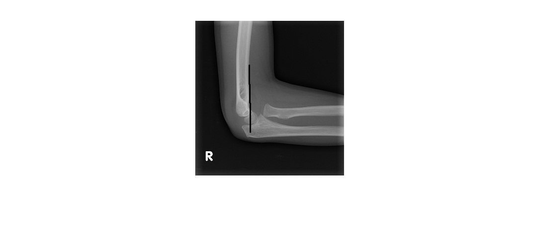
Anterior humeral line

Click to enlarge
Radiocapitellar line in AP view

Click to enlarge
Obliterated radiocapitellar line lateral view
[/peekaboo_content]