
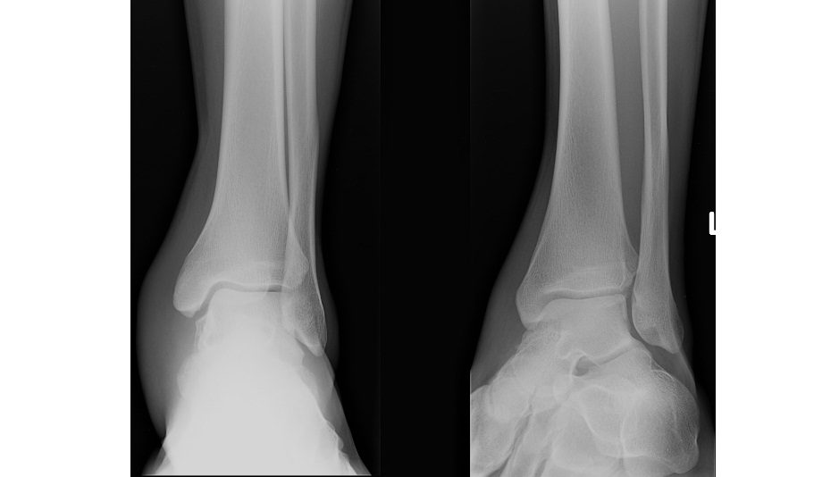
This ankle x-ray is from a 19 year old patient who inverted his ankle while running. His ankle is swollen and he is unable to weight-bear. What can you see?
Click to enlarge
All you can see is a red dot on the AP and Mortise view of the x-ray and a lot of soft tissue swelling over the medial malleolus. Can you spot the fracture?
[peekaboo_link name=”Answer”]Answer[/peekaboo_link] [peekaboo_content name=”Answer”]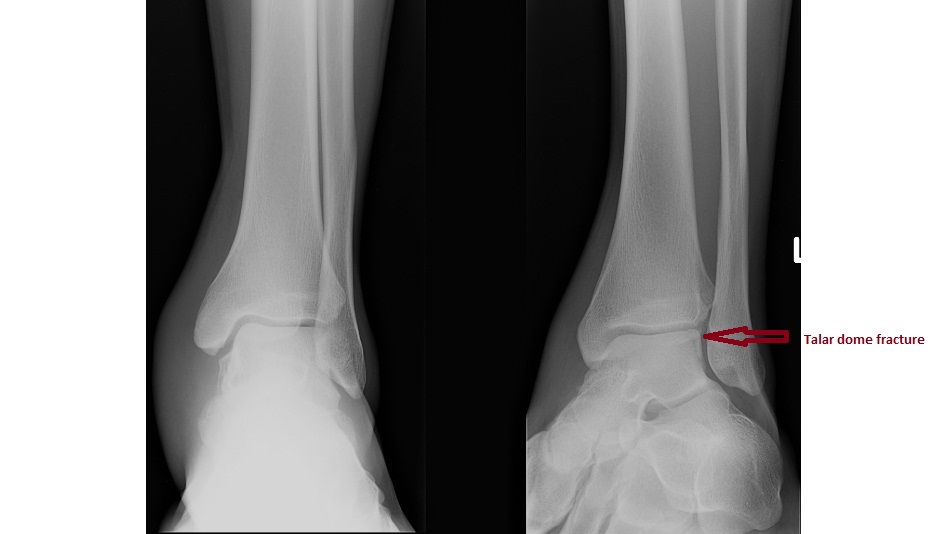
Click to enlarge
This patient has lateral talar dome fracture which is a missable fracture around the ankle.
Radiographic classification: Berndt and Harty’s
- Stage I – subchondral bone compression
- Stage II – partially detached osteochondral fragment
- Stage III – detached, nondisplaced fragment
- Stage IV – detached and displaced fragment
Treatment:
- Stage I & II – non-weight bearing short leg cast for up to 6 weeks.
- Stage III & IV – surgical management.
This patient was managed conservatively.
A mnemonic for missable fractures around the ankle (off the internet) – FLOAT
- Fifth metatarsal base
- Lateral malleolus
- Os trigonum
- Anterior process of calcaneum
- Talar dome
Hi Prathibha,
Great case.
If people want to read some more on injuries around the ankle, I’d recommend this post by @chrispartyka on his blog thebluntdissection.com.
He highlights an excellent review paper on ankle x-ray interpretation which can be found on PubMed here.
Thanks,
John
Thank you John. I must credit Dr. Parker for the case.