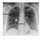
The frontal chest x-ray shows an opacified horizontal fissure as well as an increased density in the right hilum.
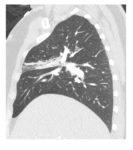
The patient had a CT pulmonary angiogram of the chest, due to clinical suspicion of a clot, which showed consolidation within the right upper lobe.The increased right hilar density is due to an enlarged hilar lymph node.
The patient was treated with oral antibiotics and a repeat chest x-ray six weeks later indicated a complete clearance of the consolidation.


Great website
Thank you.