
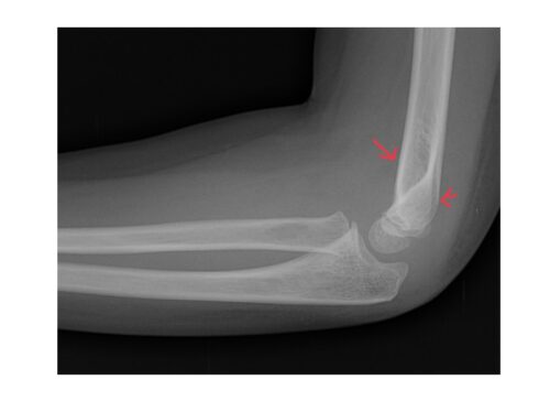
Lateral elbow x-ray shows evidence of elbow effusion with elevated fat pads (sail sign).
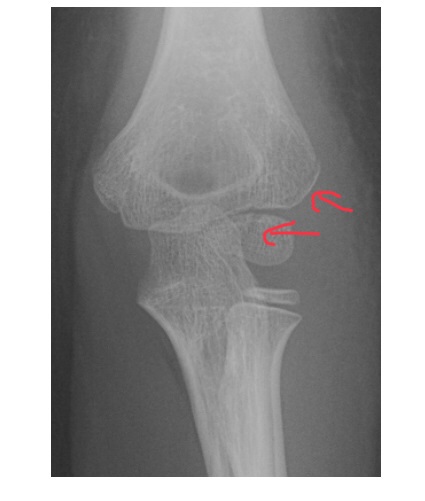
Frontal x-ray of the elbow shows minimally displaced fracture of the lateral condyle of the humerus traversing through the capitellum.
Patient was referred to the specialist and was managed conservatively.
Lateral condyle humerus fractures are the second common fractures around the elbow after supracondylar fractures in skeletally immature patients. The fracture involves the intraarticular part of the lateral condyle and can involve the non ossified cartilage of the capitellum and trochlea. The actual extent of injury seen on x-ray can be way less as a result.
These injuries should be referred to the orthopaedician from ED as operative intervention might be required.
Reference: Brant & Helms’ Fundamentals of Diagnostic Radiology, 5th Edition.