
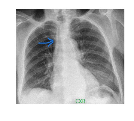
There is right paratracheal density on this rotated frontal chest x-ray.
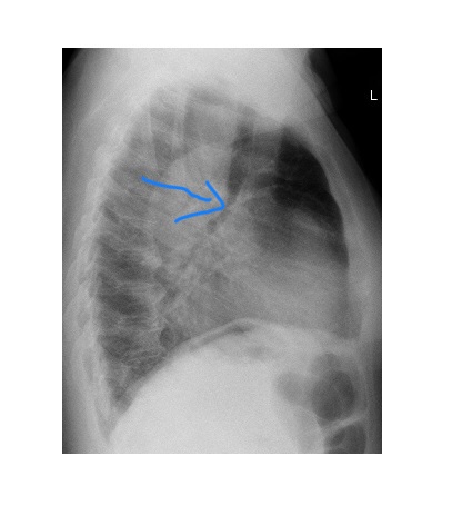
The lateral chest x-ray shows forward indentation on the posterior wall of lower trachea.
The patient underwent CT imaging of their chest which revealed significantly distended oesophagus all the way up-to the thoracic inlet.
Pay attention to the retro-tracheal area (Raider triangle) on the lateral view. Raider triangle is a radiolucent triangular space in the upper mediastinum on the lateral chest x-ray. It is bordered by the arch of aorta below, thoracic vertebral bodies posteriorly and posterior trachea anteriorly (Retrotracheal space | Radiology Reference Article | Radiopaedia.org)